. 2008 Aug;179(4):1919-32.
doi: 10.1534/genetics.108.089318. Epub 2008 Aug 9.

Regulation of apical dominance in Aspergillus nidulans hyphae by reactive oxygen species

Affiliations

Regulation of apical dominance in Aspergillus nidulans hyphae by reactive oxygen species

Camile P Semighini et al. Genetics. 2008 Aug.

Abstract

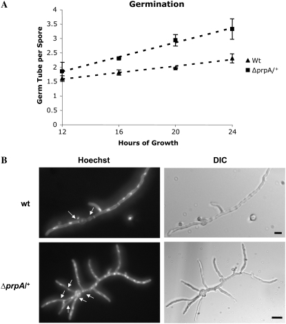
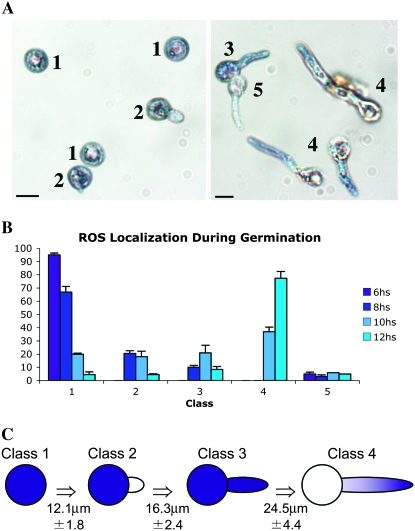
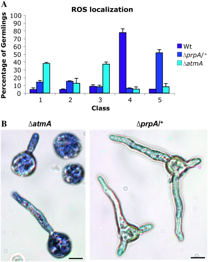
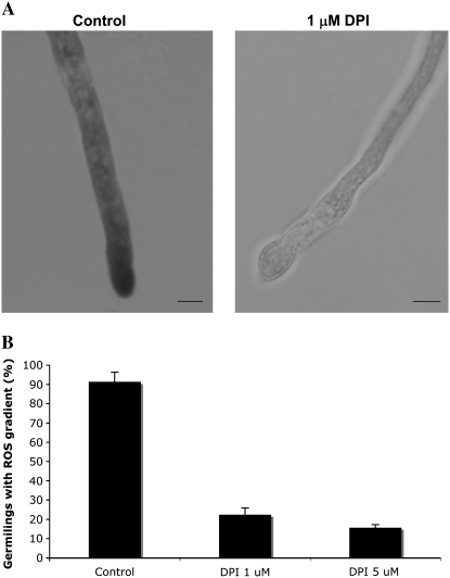
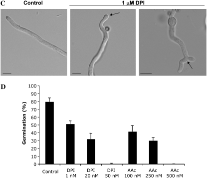
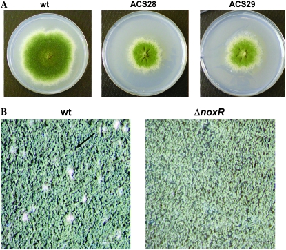
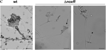
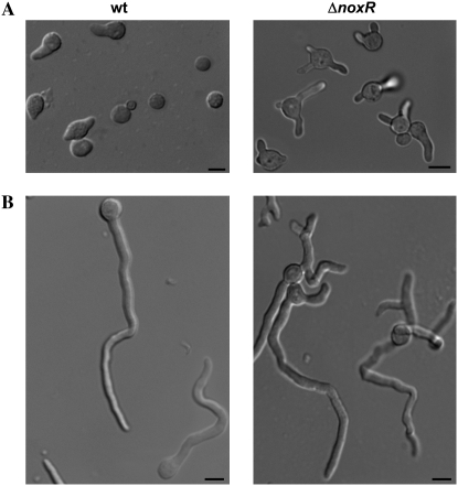
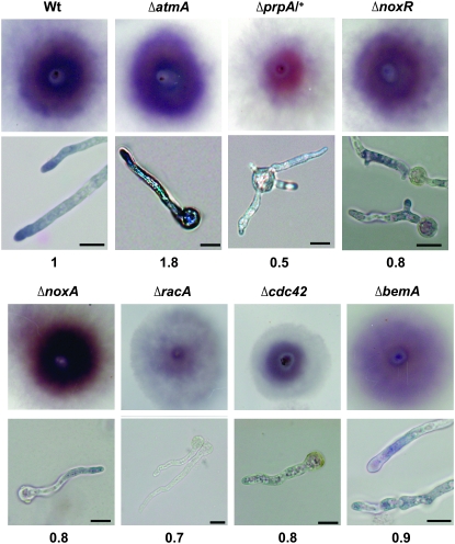
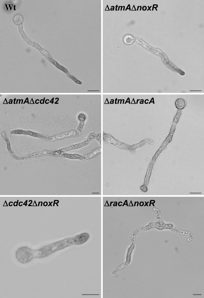
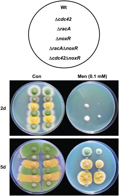
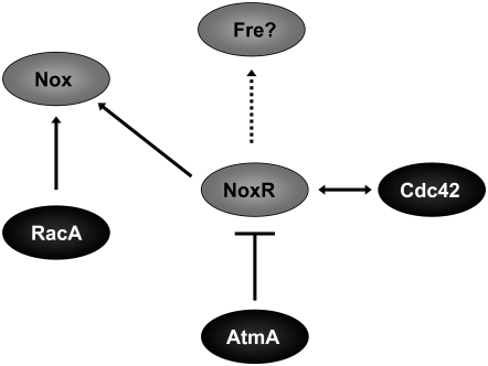
In fungal hyphae, apical dominance refers to the suppression of secondary polarity axes in the general vicinity of a growing hyphal tip. The mechanisms underlying apical dominance remain largely undefined, although calcium signaling may play a role. Here, we describe the localized accumulation of reactive oxygen species (ROS) in the apical region of Aspergillus nidulans hyphae. Our analysis of atmA (ATM) and prpA (PARP) mutants reveals a correlation between localized production of ROS and enforcement of apical dominance. We also provide evidence that NADPH oxidase (Nox) or related flavoproteins are responsible for the generation of ROS at hyphal tips and characterize the roles of the potential Nox regulators NoxR, Rac1, and Cdc42 in this process. Notably, our genetic analyses suggest that Rac1 activates Nox, whereas NoxR and Cdc42 may function together in a parallel pathway that regulates Nox localization. Moreover, the latter pathway may also include Bem1, which we propose represents a p40phox analog in fungi. Collectively, our results support a model whereby localized Nox activity generates a pool of ROS that defines a dominant polarity axis at hyphal tips.


Figures

F<sc>igure</sc> 1.—
Figure 1.—
The ΔprpA/+ mutant produces an increased number of germ tubes per spore. (A) The number of germ tubes per spore for both wild-type and ΔprpA/+ strains was determined at the indicated times. (B) Wild-type (top) and ΔprpA/+ (bottom) strains were germinated at 28° for 20 hr in YGV medium. The germinated spores were fixed and stained with Hoechst 33258 and analyzed by fluorescent microscopy. Arrows indicate germ tubes emitted from each spore. Bars, 10 μm.
F<sc>igure</sc> 2.—
Figure 2.—
ROS localization correlates to germling growth. (A) Light micrographs showing localization of ROS during conidial germination. Germlings of wild-type strain A28 grown on MN medium for 8 hr (left) and 12 hr (right) were stained with NBT. Numbers indicate the five different ROS patterns: class 1, localization throughout the entire conidium; class 2, localization in the conidium but not in the germ tube; class 3, localization throughout the entire germling; class 4, a gradient localized to the hyphal tip; and class 5, no localization. Bars, 10 μm. (B) Distribution of different patterns of ROS localization during germination. Germlings of A28 wild-type strain grown on MN medium for 6, 8, 10, and 12 hr were stained with NBT and classified into the five different ROS patterns. The graph represents the average of three independent experiments where 200 germlings were examined in each replicate. (C) Correlation between ROS localization and germling size. For each of the five patterns of ROS staining, the length of 40 germlings was determined. Average lengths are indicated with the exception of class 5, which showed no relation to hyphal size.
F<sc>igure</sc> 3.—
Figure 3.—
Disruption of ROS localization correlates with hyphal morphology defects. (A) Distribution of different patterns of ROS localization in wild-type, ΔatmA, and ΔprpA/+ strains germinated for 12 hr in MN. The graph represents the average of three independent experiments where 200 germlings were examined in each replicate. (B) Light micrographs showing localization of ROS on ΔatmA (left) and ΔprpA/+ (right) germlings grown in MN for 12 hr and stained with NBT. Bars, 10 μm.
F<sc>igure</sc> 4.—
Figure 4.—
DPI disrupts ROS gradient and induces hyphal morphology defects. (A) Light micrographs showing localization of ROS during conidial germination. Germlings of A28 wild-type strain grown on MN medium for 12 hr followed by treatment with 1 μm of DPI for 2 hr and stained with NBT. Bars, 10 μm. (B) The percentage of hyphae >25 μm containing an ROS gradient at the tip in untreated controls or following treatment with 1 or 5 μm of DPI for 2 hr. Bars show the average of three independent experiments where 200 tips were examined in each replicate. (C) Light micrographs of untreated control hyphae or hyphae treated with 1 μm of DPI for 2 hr. Arrows show abnormal hyphal-tip morphologies induced by DPI. (D) The percentage of conidia that germinated in the presence of DPI and ascorbic acid. Germlings of A28 wild-type strain were germinated on YGV medium for 16 hr in the presence of the indicated concentrations of DPI and AAc. Bars show the average of three independent experiments where 200 germlings were examined in each replicate.
F<sc>igure</sc> 5.—
Figure 5.—
Deletion of NoxR reduces colony growth and results in defective formation of conidia and cleistothecia. (A) Colony morphology of wild type and the two ΔnoxR transformants after 8 days of growth on MN at 28°. (B) Detail of colony morphology of wild-type and ΔnoxR strains after 8 days of growth on MN at 28°. Wild type forms cleistothecia (arrow) while ΔnoxR does not. Bars, 1 mm. (C) Conidiophore morphology of wild-type and ΔnoxR strains after 4 days of growth on MN at 28°. Deletion of noxR induced conidiophore defects such as branched conidiophore stalks (arrow) and the absence of vesicle plus reduced number of metulae, phialides, and conidiospores (arrowhead). Bars, 20 μm.
F<sc>igure</sc> 6.—
Figure 6.—
Deletion of NoxR results in altered germination patterns and loss of apical dominance. Germlings from wild-type (left) and ΔnoxR (right) strains grown in MN for 8 hr (A) and 12 hr (B). Bars, 10 μm. (C) Germination pattern of spores possessing two germ tubes. Spores were classified as displaying (from left to right) bipolar, quarterpolar, or random germination patterns.
F<sc>igure</sc> 7.—
Figure 7.—
ROS accumulation in candidate Nox regulators. NBT staining was used to monitor peroxide production in colonies (top) and hyphae (bottom) of the indicated mutants. Colonies were imaged following 48 hr incubation at 30° on plates. Hyphae were imaged after 16 hr incubation at 28°C on glass coverslips. Bars, 10 μm. Numbers correspond to pixel intensity of colonies stained with NBT compared to wild type (the experiment was repeated three times and similar values were found).
F<sc>igure</sc> 8.—
Figure 8.—
ROS localization in double mutants. Light micrographs showing localization of ROS in ΔatmAΔnoxR, ΔatmAΔcdc42, ΔracAΔnoxR, and Δcdc42ΔnoxR germlings grown in MN for 12 hr and stained with NBT. Bars, 10 μm.
F<sc>igure</sc> 9.—
Figure 9.—
Altered response to menadione in racA and cdc42 mutants. Approximately 103 (left column of each plate) or 102 (right column of each plate) conidia were spotted onto plates containing CM or CM plus 0.1 mm menadione. Plates were incubated at 28° and imaged after 2 and 5 days of growth.
F<sc>igure</sc> 10.—
Figure 10.—
Model for the regulatory pathways that affect Nox function. RacA is predicted to function as an activator of Nox, whereas Cdc42 and NoxR may regulate Nox localization. The dashed arrow indicates possible additional functions of NoxR, which may include, for example, the regulation of alternate NADPH oxidases such as the ferric reductase (Fre) homologs. Although the mechanism remains unknown, AtmA is predicted to negatively regulate NoxR.

References

    1. Abdallah, Y., D. Gligorievski, S. A. Kasseckert, L. Dieterich, M. Schafer et al., 2007. The role of poly(ADP-ribose) polymerase (PARP) in the autonomous proliferative response of endothelial cells to hypoxia. Cardiovasc. Res. 73 568–574. - PubMed
    1. Aguirre, J., M. Rios-Momberg, D. Hewitt and W. Hansberg, 2005. Reactive oxygen species and development in microbial eukaryotes. Trends Microbiol. 13 111–118. - PubMed
    1. Arimura, N., and K. Kaibuchi, 2005. Key regulators in neuronal polarity. Neuron 48 881–884. - PubMed
    1. Barzilai, A., and K. Yamamoto, 2004. DNA damage responses to oxidative stress. DNA Repair 3 1109–1115. - PubMed
    1. Barzilai, A., G. Rotman and Y. Shiloh, 2002. ATM deficiency and oxidative stress: a new dimension of defective response to DNA damage. DNA Repair 1 3–25. - PubMed

Publication types

MeSH terms