

doi: 10.1016/j.ddmec.2007.12.006.
Mechanisms of Post-Infarct Left Ventricular Remodeling
Affiliations
- PMID: 18690295
- PMCID: PMC2504336
- DOI: 10.1016/j.ddmec.2007.12.006
Item in Clipboard
Mechanisms of Post-Infarct Left Ventricular Remodeling
Drug Discov Today Dis Mech.
2007.
Abstract
Heart failure secondary to myocardial infarction (MI) remains a major source of morbidity and mortality. Long-term outcome after MI can be largely be defined in terms of its impact on the size and shape of the left ventricle (i.e., LV remodeling). Three major mechanisms contribute to LV remodeling: 1) early infarct expansion, 2) subsequent infarct extension into adjacent noninfarcted myocardium, and 3) late hypertrophy in the remote LV. Future developments in preventing post-MI heart failure will depend not only on identifying drugs targeting each of these individual mechanisms, but also on diagnostic techniques capable of assessing efficacy against each mechanism.
Conflict of interest statement
Figures
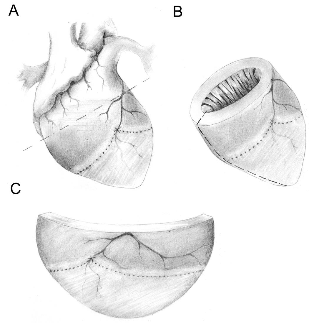
Location of the infarcted, adjacent and remote regions relative to an occluded LAD. Panel A shows entire heart with atria and major vessels. The occlusion site on the LAD is indicated with an “X”. The infarct is indicted as the lightly shaded region below the dotted line. The adjacent region is indicated as the lightly shaded region above the dotted line and the remote region is indicated as the darkly shaded region above the dotted line. Panel B shows the left ventricle with an arbitrary cut line indicated along the left-hand side as a dashed line. Panel C shows a flattened version of the LV obtained after cutting the LV along the dashed line in Panel B.
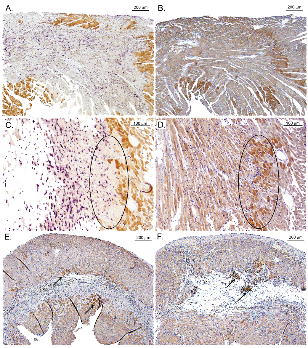
Immuno of iNOS expression in the adjacent region early and late after MI (from Gilson et al. [49] with permission). Panels A&C show low- and high-power (respectively) magnifications of a wild-type (WT) mouse heart on post-myocardial infarction (MI) day 1 double-immunostained for myoglobin (brown) and neutrophils (purple). Panels B&D show low- and high-power (respectively) serial sections from the same mouse heart immunostained for inducible nitric oxide synthase (iNOS) (brown). The iNOS immunoreactivity in panel D is most abundant in the swath of cardiomyocytes located within the black oval. Comparison with panel C reveals that the same swath of cardiomyocytes contains little myoglobin and is largely nonviable. Panels E&F show sections from WT mouse hearts on post-MI day 28 immunostained for iNOS. Panel E shows iNOS in cardiomyocytes bordering scar tissue (arrows), while Panel F shows iNOS in cardiomyocytes bordering mature scar and in granulation tissue (arrows).
References
-
- Rosamond W, et al. Heart Disease and Stroke Statistics--2007 Update: A Report From the American Heart Association Statistics Committee and Stroke Statistics Subcommittee. Circulation. 2007;115(5):e69–e171. - PubMed
-
- Mann DL. Mechanisms and models in heart failure: A combinatorial approach. Circulation. 1999;100(9):999–1008. - PubMed
-
- Mann DL, Bristow MR. Mechanisms and models in heart failure: the biomechanical model and beyond. Circulation. 2005;111(21):2837–2849. - PubMed
-
- Cohn JN. Structural basis for heart failure. Ventricular remodeling and its pharmacological inhibition. Circulation. 1995;91(10):2504–2507. - PubMed
-
- Tiyyagura SR, Pinney SP. Left ventricular remodeling after myocardial infarction: past, present, and future. Mount Sinai Journal of Medicine. 2006;73(6):840–851. - PubMed
Grants and funding
LinkOut - more resources
Full Text Sources
Other Literature Sources