Review
. 2008 Aug;9(4):338-69.
doi: 10.2174/138920308785132668.

Fluorescent proteins as biomarkers and biosensors: throwing color lights on molecular and cellular processes

Affiliations
Review

Fluorescent proteins as biomarkers and biosensors: throwing color lights on molecular and cellular processes

Olesya V Stepanenko et al. Curr Protein Pept Sci. 2008 Aug.

Abstract

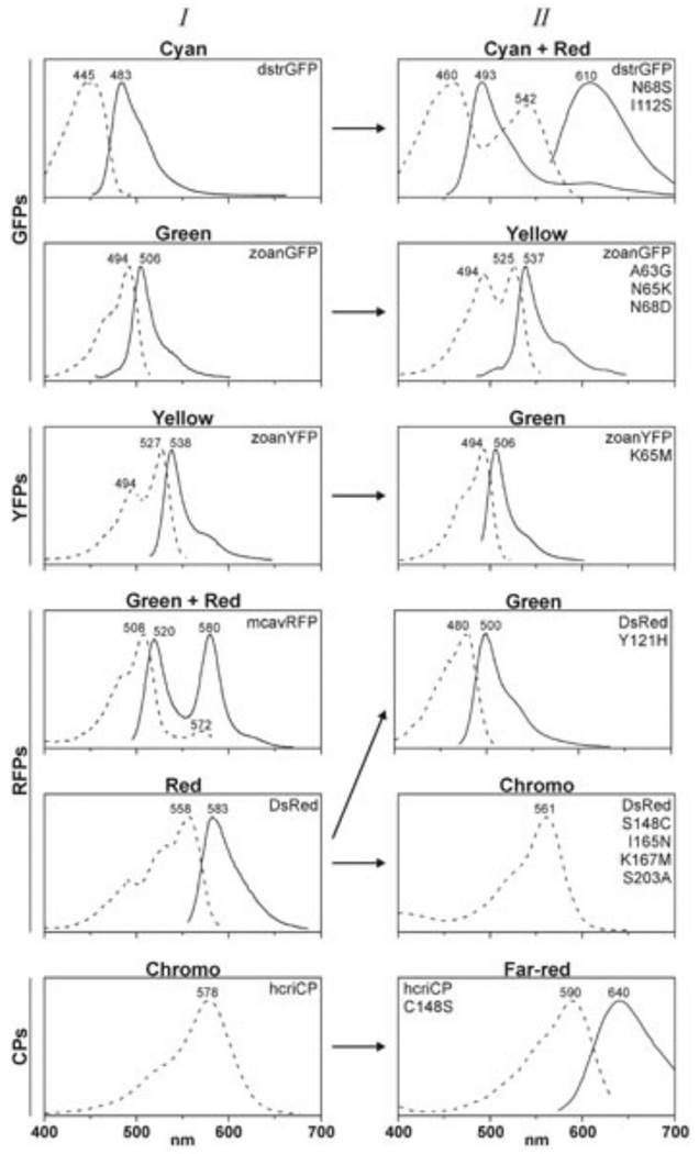
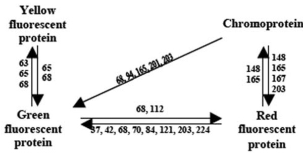
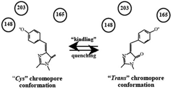
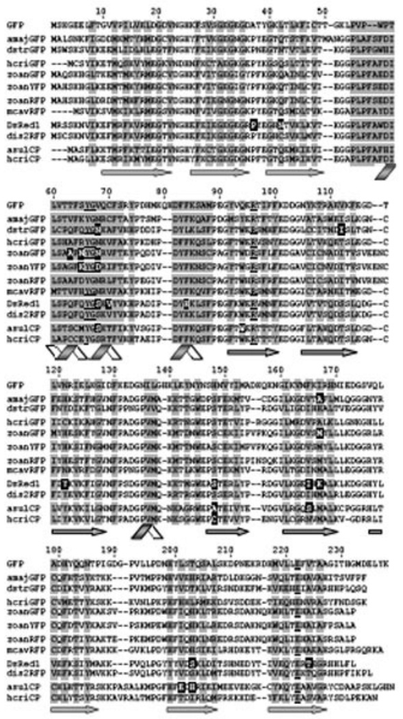
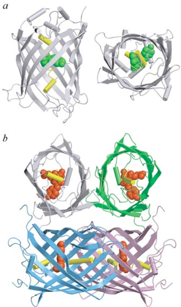
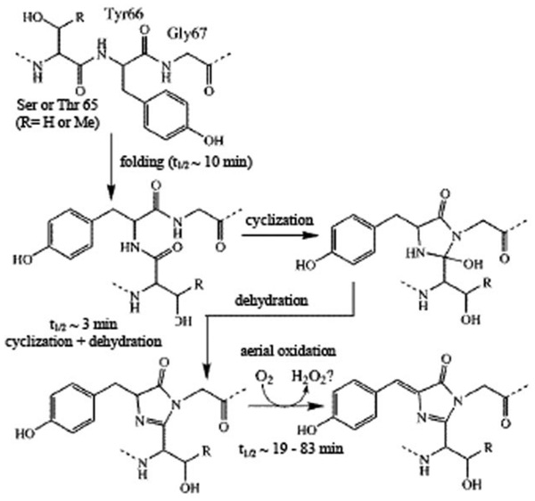
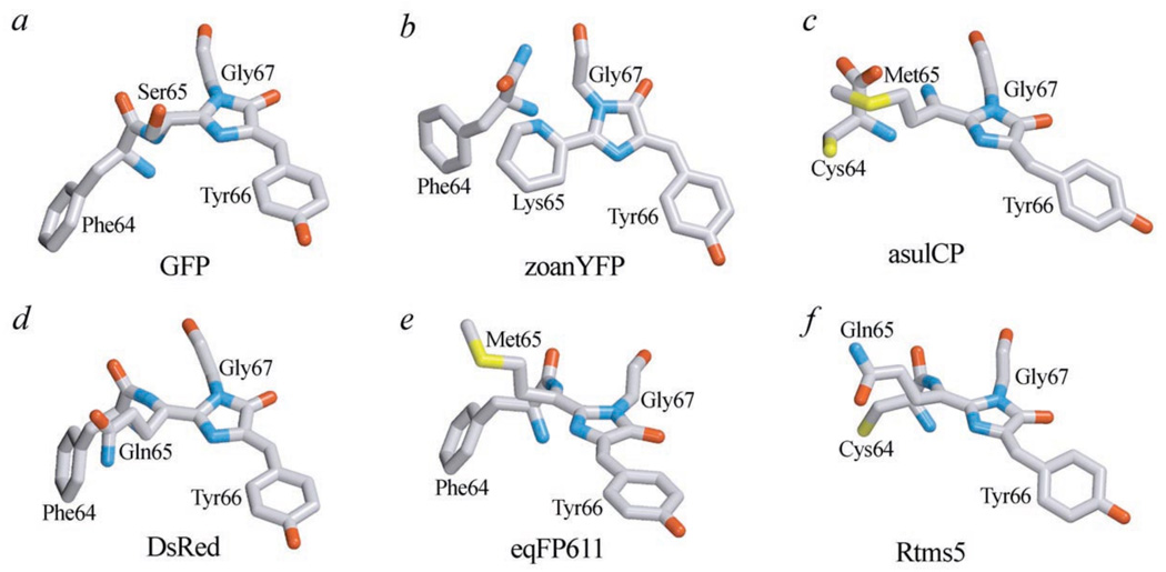
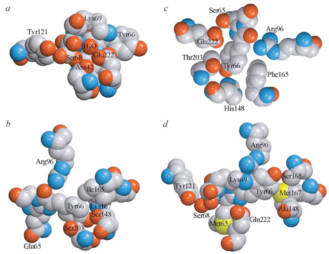
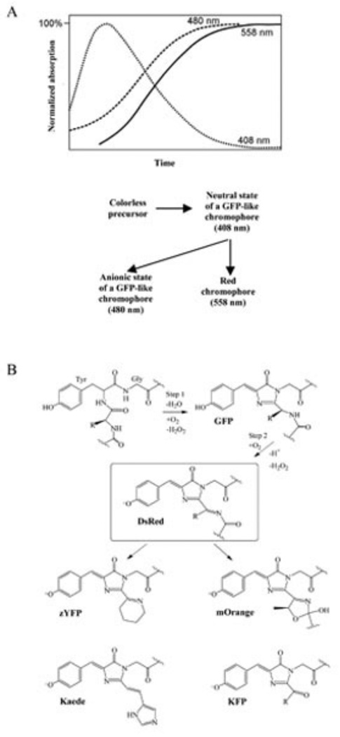
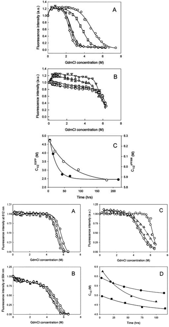
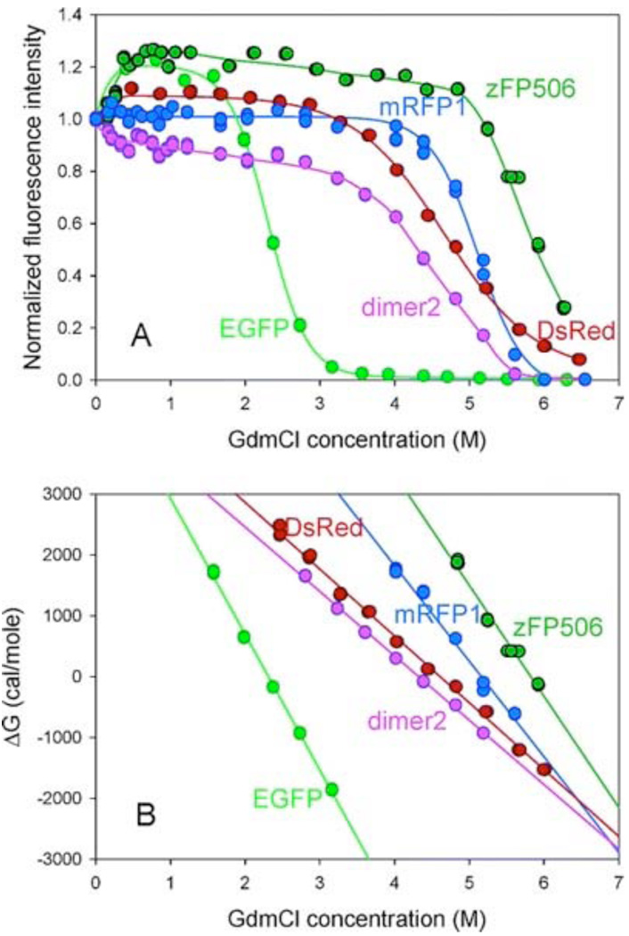
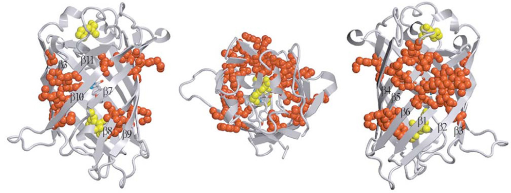
Green fluorescent protein (GFP) from jellyfish Aequorea victoria is the most extensively studied and widely used in cell biology protein. GFP-like proteins constitute a fast growing family as several naturally occurring GFP-like proteins have been discovered and enhanced mutants of Aequorea GFP have been created. These mutants differ from wild-type GFP by conformational stability, quantum yield, spectroscopic properties (positions of absorption and fluorescence spectra) and by photochemical properties. GFP-like proteins are very diverse, as they can be not only green, but also blue, orange-red, far-red, cyan, and yellow. They also can have dual-color fluorescence (e.g., green and red) or be non-fluorescent. Some of them possess kindling property, some are photoactivatable, and some are photoswitchable. This review is an attempt to characterize the main color groups of GFP-like proteins, describe their structure and mechanisms of chromophore formation, systemize data on their conformational stability and summarize the main trends of their utilization as markers and biosensors in cell and molecular biology.


Figures

Fig. (1)
Fig. (1)
Spectral properties of GFP-like proteins and their mutants belonging to the color main groups: green (GFPs), yellow (YFPs), red fluorescent proteins (RFPs) and chromoproteins (CPs) [196]. Row I – spectra of wild-type proteins, row II – spectra of mutant proteins. Dashed lines – excitation spectra of FPs or absorption spectra of CPs, solid lines – fluorescence spectra.
Fig. (2)
Fig. (2)
Amino acid substitutions responsible for the color transition of GFP-like proteins. Numbers correspond to key amino acids substituted in mutants with changed color.
Fig. (3)
Fig. (3)
Excitation and emission spectra (solid and dashed lines, respectively) and the chromophore structures of typical representatives of six classes of Aequorea GFP mutant proteins [67]. a – wild-type GFP; b – Emerald (substitutions of Ser 65 to Thr, Ser 72 to Ala, Asn 149 to Lys, Met 153 to Thr, Ile 167 to Thr), containing anionic chromophore; c – Sapphire-GFP (substitutions of Thr 203 to Ile, Ser 72 to Ala, Tyr 145 to Phe), containing neutral chromophore; d – Topaz belonging to YFPs; e – W1B from the class of CFPs; f – P4-3 belonging to BFPs.
Fig. (4)
Fig. (4)
Cis–trans isomerization of KFP1 chromophore during reversible kindling and quenching. Numbers – the amino acids stabilizing the chromophore in fluorescent or non-fluorescent state.
Fig. (5)
Fig. (5)
Amino acid sequences of GFP-like proteins possessing various fluorescent properties: amajGFP and dstrGFP – subgroup of cyan fluorescent proteins, hcriGFP and zoanGFP – green fluorescent proteins, zoanYFP – yellow fluorescent proteins, DsRed1 and dis2RFP – red fluorescent proteins, and asulCP and hcriCP – non-fluorescent proteins or chromoproteins. ZoanRFP and mcavRFP show dual-color fluorescence [196]. Numbering of amino acids corresponds to Aequorea GFP. Conserved Tyr 66, Gly 67, Arg 96 and Glu 222 are underlined. Shaded areas point out the amino acids whose side chains form the interior of the β-barrel. Amino acids, which are crucial for fluorescent properties formation, are displayed in white on black. Figures below the sequences show the elements of secondary structure.
Fig. (6)
Fig. (6)
X-ray crystal structure of Aequorea GFP (PDB code 1W7S) in two projections (a) and of DsRed1 from Discosoma sp. (PDB code 1G7K) (b). Chromophores of GFP and DsRed1 are shown as green and red space-filling unions, respectively. A central α-helix which includes chromophore is shown in yellow. Monomers of DsRed1 are displayed in different colors. The drawing was generated by the graphic programs VMD [197] and Raster3D [198, 199].
Fig. (7)
Fig. (7)
Mechanism of GFP chromophore formation [19]. Rate constants were estimated for mutant of GFP containing the substitution of Ser 65 to Thr [20, 200].
Fig. (8)
Fig. (8)
Different types of chromophore in GFP-like proteins. a –GFP from Aequorea victoria (PDB code 1W7S); b – yellow fluorescent protein zoanYFP from Zoanthus sp. (PDB code 1XAE); c - red fluorescent protein DsRed from Discosoma sp. (PDB code 1G7K); d – far-red fluorescent protein eqFP611 from Entacmaea (PDB code 1UIS); e – non-fluorescent chromoprotein asulCP from Anemonia sulcata (PDB code 2A50); f – non-fluorescent chromoprotein Rtms5 from Montipora (PDB code 1MOU). Numbering of amino acids corresponds to Aequorea GFP. The drawing was generated by the graphic programs VMD [197] and Raster3D [198, 199].
Fig. (9)
Fig. (9)
The microenvironment of DsRed1 chromophore (a and b), Aequorea GFP chromophore (c) and asulCP chromophore (d). Carbon, nitrogen, oxygen and sulfur are gray blue, red and yellow, respectively. The drawing was generated by graphic programs VMD [197] and Raster3D [, .].
Fig. 10
Fig. 10
(A) Time course of the formation of blue, green and red forms of the DsRed1 chromophore with absorption maxima at 408, 480 and 558 nm, respectively (top) and proposed scheme for these spectral forms formation from non-fluorescent precursor (bottom) [51]. (B) The schematic diagram of the chromophore formation in various GFP-like proteins. The GFP- and DsRed-like chromophores are supposed to be intermediates on the pathway of some other chromophore maturation [57].
Fig (11)
Fig (11)
Quasi-equilibrium unfolding of EGFP (A), zFP506 (B), mRFP1 (C), "dimer2" (D) and DsRed (E) induced by GdmCl. Measurements for EGFP were performed after 5 (circles), 15 (squares), 43 (triangles), 67 (reversed triangles) and 216 hrs (diamonds) of incubation in the presence of desired GdmCl concentration. Fluorescence was excited at excited at 365 nm and detected at 510 nm for EGFP and zFP506, 585 nm for DsRed1 and "dimer2", and at 610 nm for mRFP1. Measurements for zFP506 were done after 1 (circles), 2 (squares), 3 (triangles) and 5 days (reversed triangles) of incubation in the presence of desired GdmCl concentration. Measurements for mRFP1 were performed after 1 (circles), 2 (squares), 3 (triangles), and 5 days (diamonds) of incubation in the presence of desired GdmCl concentration. Measurements for "dimer2" were done after 1 (circles), 2 (squares), 3 (triangles) and 5 days (dimonds) of incubation in the presence of desired GdmCl concentration. Fluorescence was excited at excited at 365 nm and detected at 510 nm for EGFP and zFP506, 585 nm for DsRed1 and "dimer2", and at 610 nm for mRFP1. Measurements for DsRed were done after 5 (circles), 24 (squares), 56 (triangles), 100 hrs (diamonds) of incubation in the presence of desired GdmCl concentration.
Fig. (12)
Fig. (12)
Equilibrium GdmCl-induced unfolding of EGFP (light green circles and lines) zFP506 (dark green circles and lines), mRFP1 (blue circles and lines) "dimer2" (pink circles and lines), and DsRed (red circles and lines) detected by GdmCl-induced changes in characteristic green or red fluorescence measured after the 9 days of incubation in the presence of desired GdmCl concentration.
Fig. (13)
Fig. (13)
Three-dimensional representation of the positions of residues in GFPuv (PDB code 1B9C) with very slow H/D exchange rate constants for the amide groups [74]. Left and right figures are viewed from two opposite sides. The central figure is from the top of the β-barrel. The chromophore is shown in stick mode. Red and yellow balls represent very slow exchanging residues in β-strands and α-helixes, respectively. Each β-strand is numbered from the N to the C terminus. The drawing was generated by the graphic programs VMD [197] and Raster3D [198, 199].
Fig. (14)
Fig. (14)
Possible topologies of GFP (cylinders), circularly permuted GFP (cpGFP, cylinders) and chimeras with other proteins (spheres and hemisphers) [191]. Figure depicts non-modified GFP (a), tandem fusion of a target protein to GFP (b), GFP insertion into the target protein (c), insertion of a target protein into cpGFP (d), cpGFP (e), fusion of a target protein to cpGFP (f), cpGFP insertion into the target protein (g), and insertion of a target protein into GFP (h).

References

    1. Labas YA, Gurskaya NG, Yanushevich YG, Fradkov AF, Lukyanov KA, Lukyanov SA, Matz MV. Proc Natl Acad Sci U S A. 2002;99:4256–4261. - PMC - PubMed
    1. Lukyanov KA, Fradkov AF, Gurskaya NG, Matz MV, Labas YA, Savitsky AP, Markelov ML, Zaraisky AG, Zhao X, Fang Y, Tan W, Lukyanov SA. J Biol Chem. 2000;275:25879–25882. - PubMed
    1. Cubitt AB, Heim R, Adams SR, Boyd AE, Gross LA, Tsien RY. Trends Biochem Sci. 1995;20:448–455. - PubMed
    1. Zacharias DA, Baird GS, Tsien RY. Curr Opin Neurobiol. 2000;10:416–421. - PubMed
    1. Ward WW, Cormier MJ. J Biol Chem. 1979;254:781–788. - PubMed

Publication types

LinkOut - more resources