

Functional implications for Kir4.1 channels in glial biology: from K+ buffering to cell differentiation
- PMID: 18691387
- PMCID: PMC2581639
- DOI: 10.1111/j.1471-4159.2008.05615.x
Functional implications for Kir4.1 channels in glial biology: from K+ buffering to cell differentiation
Abstract
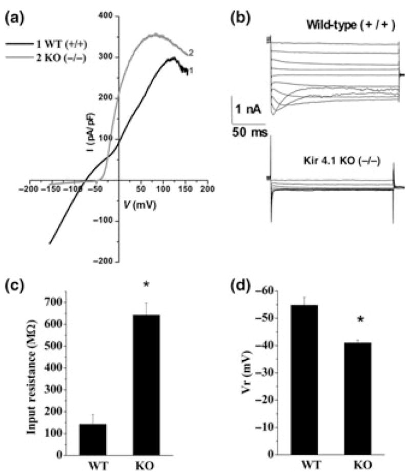
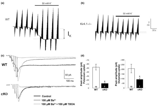
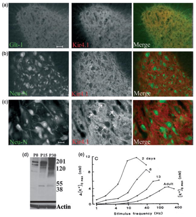
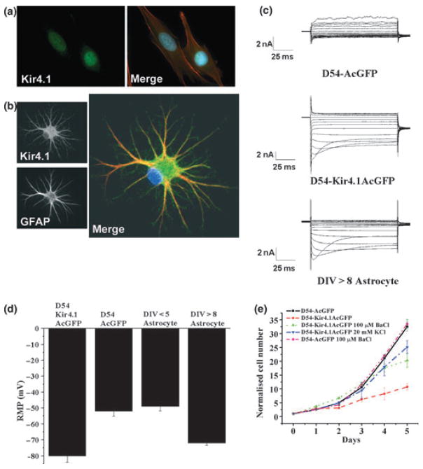
Astrocytes and oligodendrocytes are characterized by a very negative resting potential and a high resting permeability for K(+) ions. Early pharmacological and biophysical studies suggested that the resting potential is established by the activity of inwardly rectifying, Ba(2+) sensitive, weakly rectifying Kir channels. Molecular cloning has identified 16 Kir channels genes of which several mRNA transcripts and protein products have been identified in glial cells. However, genetic deletion and siRNA knock-down studies suggest that the resting conductance of astrocytes and oligodendrocytes is largely due to Kir4.1. Loss of Kir4.1 causes membrane depolarization, and a break-down of K(+) and glutamate homeostasis which results in seizures and wide-spread white matter pathology. Kir channels have also been shown to act as critical regulators of cell division whereby Kir function is correlated with an exit from the cell cycle. Conversely, loss of functional Kir channels is associated with re-entry of cells into the cell cycle and gliosis. A loss of functional Kir channels has been shown in a number of neurological diseases including temporal lobe epilepsy, amyotrophic lateral sclerosis, retinal degeneration and malignant gliomas. In the latter, expression of Kir4.1 is sufficient to arrest the aberrant growth of these glial derived tumor cells. Kir4.1 therefore represents a potential therapeutic target in a wide variety of neurological conditions.
Figures

Similar articles
-
Inwardly rectifying potassium channels (Kir) in central nervous system glia: a special role for Kir4.1 in glial functions.J Cell Mol Med. 2006 Jan-Mar;10(1):33-44. doi: 10.1111/j.1582-4934.2006.tb00289.x. J Cell Mol Med. 2006. PMID: 16563220 Free PMC article. Review.
-
Kir potassium channel subunit expression in retinal glial cells: implications for spatial potassium buffering.Glia. 2002 Sep;39(3):292-303. doi: 10.1002/glia.10112. Glia. 2002. PMID: 12203395
-
Potassium channel activity and glutamate uptake are impaired in astrocytes of seizure-susceptible DBA/2 mice.Epilepsia. 2010 Sep;51(9):1707-13. doi: 10.1111/j.1528-1167.2010.02592.x. Epilepsia. 2010. PMID: 20831751 Free PMC article.
-
Conditional knock-out of Kir4.1 leads to glial membrane depolarization, inhibition of potassium and glutamate uptake, and enhanced short-term synaptic potentiation.J Neurosci. 2007 Oct 17;27(42):11354-65. doi: 10.1523/JNEUROSCI.0723-07.2007. J Neurosci. 2007. PMID: 17942730 Free PMC article.
-
The role of an inwardly rectifying K(+) channel (Kir4.1) in the inner ear and hearing loss.Neuroscience. 2014 Apr 18;265:137-46. doi: 10.1016/j.neuroscience.2014.01.036. Epub 2014 Jan 28. Neuroscience. 2014. PMID: 24480364 Free PMC article. Review.
Cited by
-
Human glioma-initiating cells show a distinct immature phenotype resembling but not identical to NG2 glia.J Neuropathol Exp Neurol. 2013 Apr;72(4):307-24. doi: 10.1097/NEN.0b013e31828afdbd. J Neuropathol Exp Neurol. 2013. PMID: 23481707 Free PMC article.
-
Antidepressant mechanisms of ketamine: a review of actions with relevance to treatment-resistance and neuroprogression.Front Neurosci. 2023 Aug 8;17:1223145. doi: 10.3389/fnins.2023.1223145. eCollection 2023. Front Neurosci. 2023. PMID: 37614344 Free PMC article. Review.
-
Deeper and Deeper on the Role of BK and Kir4.1 Channels in Glioblastoma Invasiveness: A Novel Summative Mechanism?Front Neurosci. 2020 Nov 30;14:595664. doi: 10.3389/fnins.2020.595664. eCollection 2020. Front Neurosci. 2020. PMID: 33328867 Free PMC article. Review.
-
Potassium Current Signature of Neuronal/Glial Progenitors in Amniotic Fluid Stem Cells.Cells. 2025 Jan 4;14(1):50. doi: 10.3390/cells14010050. Cells. 2025. PMID: 39791751 Free PMC article.
-
Preclinical Insights into the Role of Kir4.1 in Chronic Pain and Depression: Mechanisms and Therapeutic Potential.Biomolecules. 2025 Jan 23;15(2):165. doi: 10.3390/biom15020165. Biomolecules. 2025. PMID: 40001468 Free PMC article. Review.
References
-
- Amiry-Moghaddam M, Williamson A, Palomba M, Eid T, de Lanerolle NC, Nagelhus EA, Adams ME, Froehner SC, Agre P, Ottersen OP. Delayed K+ clearance associated with aquaporin-4 mislocalization: phenotypic defects in brains of alpha-syntrophin-null mice. Proc Natl Acad Sci USA. 2003;100:13615–13620. - PMC - PubMed
-
- Anderson CM, Swanson RA. Astrocyte glutamate transport: review of properties, regulation, and physiological functions. Glia. 2000;32:1–14. - PubMed
-
- Benfenati V, Caprini M, Nobile M, Rapisarda C, Ferroni S. Guanosine promotes the up-regulation of inward rectifier potassium current mediated by Kir4.1 in cultured rat cortical astrocytes. J Neurochem. 2006;98:430–445. - PubMed
-
- Bordey A, Sontheimer H. Postnatal development of ionic currents in rat hippocampal astrocytes in situ. J Neurophysiol. 1997;78:461–477. - PubMed
Publication types
MeSH terms
Substances
Grants and funding
LinkOut - more resources
Full Text Sources
Other Literature Sources
Medical
