

Canine vector-borne diseases in Brazil
- PMID: 18691408
- PMCID: PMC2533296
- DOI: 10.1186/1756-3305-1-25
Canine vector-borne diseases in Brazil
Abstract
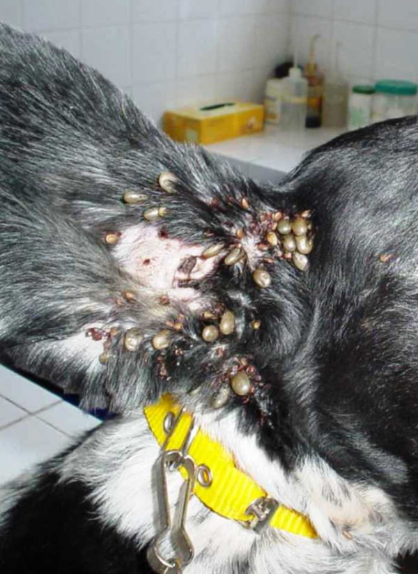
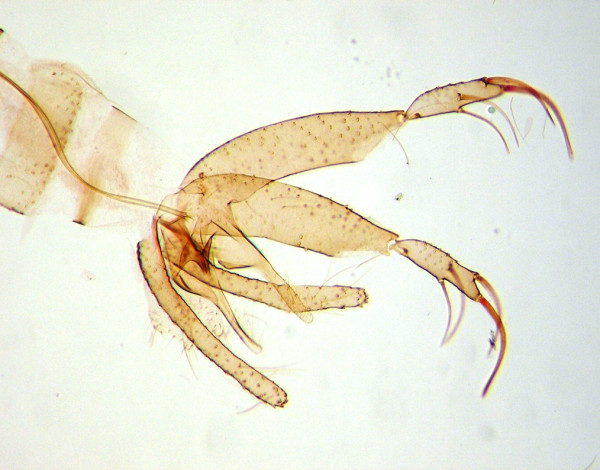
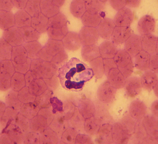
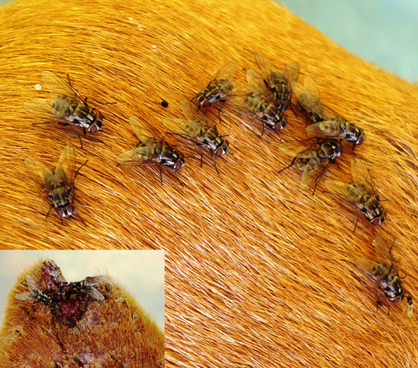
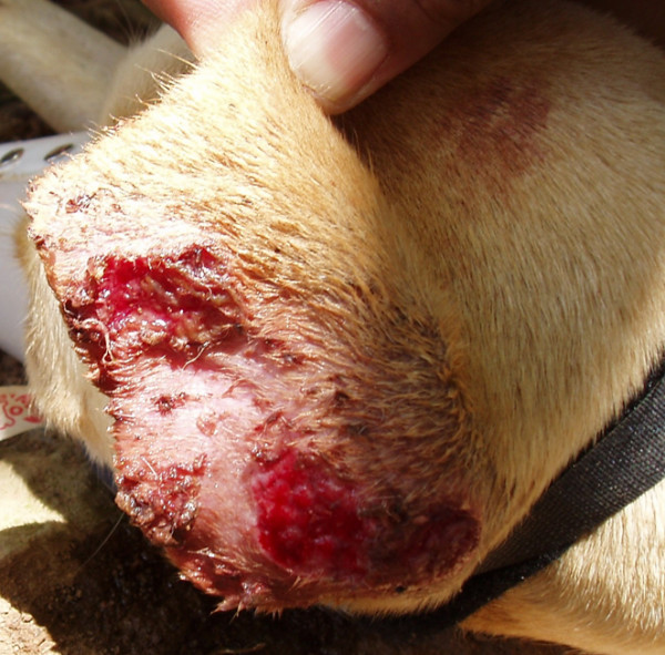
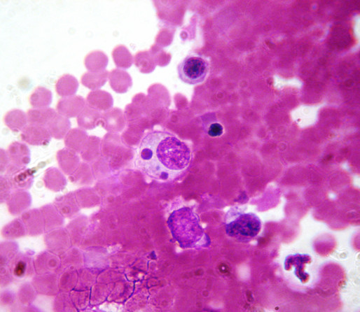
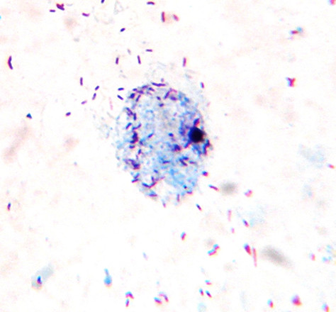
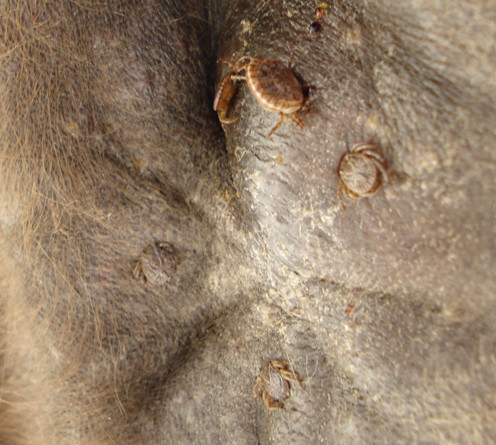
Canine vector-borne diseases (CVBDs) are highly prevalent in Brazil and represent a challenge to veterinarians and public health workers, since some diseases are of great zoonotic potential. Dogs are affected by many protozoa (e.g., Babesia vogeli, Leishmania infantum, and Trypanosoma cruzi), bacteria (e.g., Anaplasma platys and Ehrlichia canis), and helminths (e.g., Dirofilaria immitis and Dipylidium caninum) that are transmitted by a diverse range of arthropod vectors, including ticks, fleas, lice, triatomines, mosquitoes, tabanids, and phlebotomine sand flies. This article focuses on several aspects (etiology, transmission, distribution, prevalence, risk factors, diagnosis, control, prevention, and public health significance) of CVBDs in Brazil and discusses research gaps to be addressed in future studies.
Figures


References
-
- Irwin PJ. Companion animal parasitology: a clinical perspective. Int J Parasitol. 2002;32:581–593. - PubMed
-
- Hunter PR. Climate change and waterborne and vector-borne disease. J Appl Microbiol. 2003;94:37S–46S. - PubMed
-
- Silva-Araújo A. Filaria immitis e a Filaria sanguinolenta no Brasil. Gaz Méd Bahia. 1878;7:295–312.
-
- Camargo-Neves VL, Katz G, Rodas LA, Poletto DW, Lage LC, Spínola RM, Cruz OG. Utilização de ferramentas de análise espacial na vigilância epidemiológica de leishmaniose visceral americana – Araçatuba, São Paulo, Brasil, 1998–1999. Cad Saúde Pública. 2001;17:1263–1267. - PubMed
-
- Savani ESMM, Schimonsky B, Camargo MCGO, D'auria SRN. Vigilância de leishmaniose visceral americana em cães de área não endêmica, São Paulo. Rev Saúde Publica. 2003;37:260–262. - PubMed
LinkOut - more resources
Full Text Sources
