. 2008 Aug 8:5:33.
doi: 10.1186/1742-2094-5-33.

P2x7 deficiency suppresses development of experimental autoimmune encephalomyelitis

Affiliations

P2x7 deficiency suppresses development of experimental autoimmune encephalomyelitis

Anthony J Sharp et al. J Neuroinflammation. .

Abstract

Background: The purinergic receptor P2x7 is expressed on myeloid cells as well as on CNS glial cells, and P2x7 activation has been shown to increase both glial and T-cell activation. These properties suggest a role in the development of autoimmune disease including multiple sclerosis.

Methods: The animal model of MS, experimental autoimmune encephalomyelitis (EAE) using myelin oligodendrocyte glycoprotein (MOG) peptide residues 35-55 was induced in wildtype C57BL6 mice and in P2x7 deficient mice ('P2x7 mice') that were backcrossed to C57BL6 mice. Disease progression was monitored by appearance of clinical signs, immunocytochemical staining to assess brain inflammation and neuronal damage, and by measurement of Tcell cytokine production.

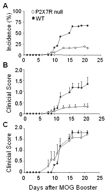
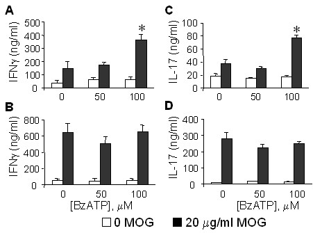
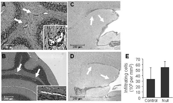
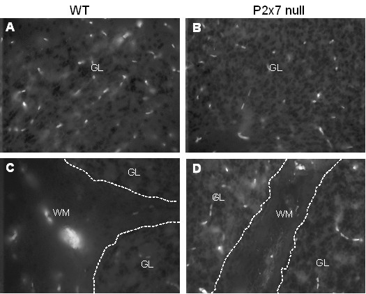
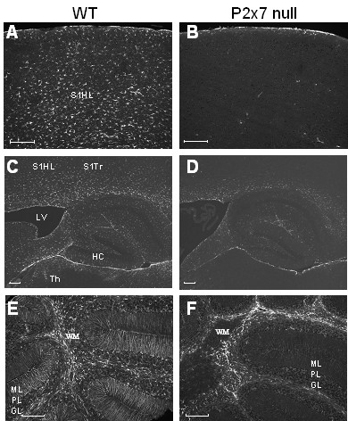
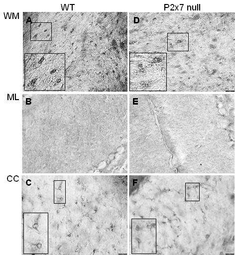
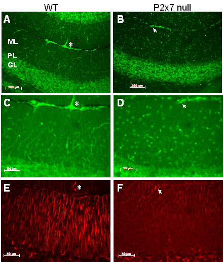
Results: The incidence of EAE disease in P2x7 mice was reduced 4-fold compared to the wildtype mice; however the P2x7 mice that became ill had similar days of onset and clinical scores as the wildtype mice. Splenic T-cells isolated from P2x7 null mice produced greater IFNgamma and IL-17 (from 3 to 12 fold greater levels) than wildtype cells, however cytokine production from P2x7 derived cells was not increased by a selective P2x7 agonist as was cytokine production from wildtype cells. Although infiltrating cells were detected in brains of both the P2x7 and wildtype mice, astroglial activation and axonal damage was reduced versus wildtype mice, and the distribution of astroglial activation was markedly distinct in the two strains. In contrast, microglial activation was similar in the two strains.

Conclusion: P2x7 deficiency resulted in compensatory changes leading to increased T-cell cytokine production, and activated T-cells were detected in the brains of P2x7 null mice with no clinical signs. However, the greatly reduced incidence of disease suggests that an initiating event is absent in these mice, and points to a role for astroglial P2x7 in development of EAE disease.


Figures

Figure 1
Figure 1
Effect of P2x7 deficiency on the development of clinical signs of EAE. P2x7 null and wildtype C57BL6 mice were immunized to develop EAE using MOG35–55 peptide, and clinical scores monitored approximately daily. Day 0 is defined as the day of the MOG peptide booster immunization. The data from 3 separate studies using P2x7 null mice (n = 38 total) and 2 separate studies using WT mice (n = 24 total) are combined. Data in (A) shows average incidence of disease (defined by at least 2 consecutive days with a score of 1.0 or greater). Data in (B) shows average clinical scores (mean ± se) for all mice. Data in (C) show average clinical scores (mean ± se) for only those mice that became ill (7/38 in the P2x7 null group; and 15/24 in the WT group).
Figure 2
Figure 2
Effect of P2x7 deficiency on splenic T-cell cytokine production. P2x7 and wildtype C57BL6 mice were immunized to develop EAE as above, and at 4 days after the booster immunization, splenic T-cells isolated and cultured in the presence (filled bars) or absence (open bars) of 20 μg/ml MOG peptide, and either 0, 50, or 100 μM BzATP. After 24 hr, levels of IFNγ and IL-17 in the culture media were determined by specific ELISA. Cytokine production is shown for WT (A, C) and P2x7 null (B, D) cells. Values are the mean ± sd of n = 3 independent samples. *, P < .001 versus no BzATP.
Figure 3
Figure 3
Histological staining for infiltrating cells in P2x7 and WT mice. Serial sections were prepared from EAE immunized P2x7 null and WT mice at day 21 after immunization. At this time, the 3 WT mice had clinical scores of 2.0; and the 3 P2x7 null mice had no clinical signs. Sections were stained with H&E to detect the presence of infiltrating cells. Representative sections from the cerebellum show the presence of perivascular infiltrating cells (arrows) in the cerebellar white matter of both P2x7 (A) and WT (B) brains. Sections taken from near the IVth ventricle show the presence of infiltrating cells (arrows) beneath the corpus callosum and lining the ventricle in both P2x7 (C) and WT (D) brains. (E) Quantitation of the average total number of infiltrating cells (mean ± sd) measured in 4 serial sections for P2x7 and WT mice (n = 3 mice each).
Figure 4
Figure 4
CD8 T-cells are expressed in WT and P2x7 cerebellum. Serial sections as prepared in Figure 3 were stained with antibody to CD8 to detect the presence of T-cells. Representative images from one mouse in each group show the presence of CD8 cells in both the granule layer ('GL', Panels A and B) and white matter ('WM', Panels C and D) of WT and P2x7 animals.
Figure 5
Figure 5
GFAP expression is reduced in P2x7 brain. Serial sections were prepared from EAE immunized P2x7 null and WT mice at day 21 after immunization. At this time, the 3 WT mice all had clinical signs of 2.0; and the 3 P2x7 null mice had no clinical signs. Sections were stained with antibody to GFAP to detect activated astrocytes. Representative images show robust GFAP staining in WT frontal cortex (A) and thalamus (C), but not in corresponding areas of P2x7 null cortex (B) or thalamus (D). Significant staining for GFAP was found throughout the cerebellar grey and white matter in the WT mice (E) but was restricted to white matter (W) in P2x7 mice (F). S1HL, somatosensory hindlimb region; S1Trl, somatosensory trunk region; LV, lateral ventricle; HC, hippocampus; Th, laterodorsal thalamic nuclei; WM, white matter; ML, molecular layer; PL, Purkinje cell layer; GL, granule cell layer. Size bars are 200 μm.
Figure 6
Figure 6
Microglial activation is similar in P2x7 and WT brain. Serial sections from WT (A, B, C) and P2x7 null (E, F, G) mice as described in Figure 5 were stained with monoclonal antibody F4/80 to detect activated microglia and macrophages. Representative images from one mouse in each group show the presence of cells with characteristic morphology of microglia in cerebellar white matter ('WM', panels A, D) and corpus callosum ('CC', panels C, F); and diffuse staining in cerebellar granule layer ('GL', panels B, E).
Figure 7
Figure 7
Axonal damage is reduced in P2x7 brain. Serial sections were prepared from EAE immunized P2x7 null and WT mice at day 21 after immunization. At this time, the 3 WT mice all had clinical signs of 2.0; and the 3 P2x7 null mice had no clinical signs. Sections were stained with antibody SMI32 (for non phosphorylated neurofilament proteins, A-D) or to GFAP (E, F). Representative images show significant SMI32 staining in an area of the cerebellum from WT (A, C) mice, but little staining in a comparable area from P2x7 null (B, D) mice. Size bars are 100 μM (A, B) and 50 μm (C-F). GFAP staining (E, F) illustrates the differences in localization for GFAP and SMI32 staining. Asterisks and arrows are placed to indicate same location in images. ML, molecular layer; PL, Purkinje cell layer; GL, granule cell layer.

References

    1. Narcisse L, Scemes E, Zhao Y, Lee SC, Brosnan CF. The cytokine IL-1beta transiently enhances P2X7 receptor expression and function in human astrocytes. Glia. 2005;49:245–258. doi: 10.1002/glia.20110. - DOI - PMC - PubMed
    1. Marcoli M, Cervetto C, Paluzzi P, Guarnieri S, Alloisio S, Thellung S, Nobile M, Maura G. P2X(7) pre-synaptic receptors in adult rat cerebrocortical nerve terminals: a role in ATP-induced glutamate release. J Neurochem. 2008 - PubMed
    1. Hamilton N, Vayro S, Kirchhoff F, Verkhratsky A, Robbins J, Gorecki DC, Butt AM. Mechanisms of ATP- and glutamate-mediated calcium signaling in white matter astrocytes. Glia. 2008;56:734–749. doi: 10.1002/glia.20649. - DOI - PubMed
    1. Matute C, Torre I, Perez-Cerda F, Perez-Samartin A, Alberdi E, Etxebarria E, Arranz AM, Ravid R, Rodriguez-Antiguedad A, Sanchez-Gomez M, Domercq M. P2X(7) receptor blockade prevents ATP excitotoxicity in oligodendrocytes and ameliorates experimental autoimmune encephalomyelitis. J Neurosci. 2007;27:9525–9533. doi: 10.1523/JNEUROSCI.0579-07.2007. - DOI - PMC - PubMed
    1. Agresti C, Meomartini ME, Amadio S, Ambrosini E, Volonte C, Aloisi F, Visentin S. ATP regulates oligodendrocyte progenitor migration, proliferation, and differentiation: involvement of metabotropic P2 receptors. Brain Res Brain Res Rev. 2005;48:157–165. doi: 10.1016/j.brainresrev.2004.12.005. - DOI - PubMed

Publication types

MeSH terms

LinkOut - more resources