. 2008 Sep 26:1232:70-84.
doi: 10.1016/j.brainres.2008.07.058. Epub 2008 Jul 26.

Ovarian steroids modulate leu-enkephalin levels and target leu-enkephalinergic profiles in the female hippocampal mossy fiber pathway

Affiliations

Ovarian steroids modulate leu-enkephalin levels and target leu-enkephalinergic profiles in the female hippocampal mossy fiber pathway

Annelyn Torres-Reveron et al. Brain Res. .

Abstract

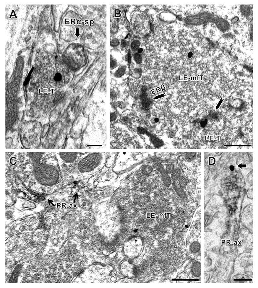
In the hippocampal formation (HF), the enkephalin opioids and estrogen are each known to modulate learning and cognitive performance relevant to drug abuse. Within the HF, leu-enkephalin (LENK) is most prominent in the mossy fiber (MF) pathway formed by the axons of dentate gyrus (DG) granule cells. To examine the influence of ovarian steroids on MF pathway LENK levels, we used quantitative light microscopic immunocytochemistry to evaluate LENK levels in normal cycling rats and in estrogen-treated ovariectomized rats. Rats in estrus had increased levels of LENK-immunoreactivity (ir) in the DG hilus compared to rats in diestrus or proestrus. Rats in estrus and proestrus had higher levels of LENK-ir in CA3a-c compared to rats in diestrus. Ovariectomized (OVX) rats 24 h (but not 6 or 72 h) after estradiol benzoate (EB; 10 microg) administration had increased LENK-ir in the DG hilus and CA3c. Electron microscopy showed a larger proportion of LENK-labeled small terminals and axons in the DG hilus compared to CA3 which may have contributed to region-specific changes in LENK-ir densities. Next we evaluated the subcellular relationships of estrogen receptor (ER) alpha, ERbeta and progestin receptor (PR) with LENK-labeled MF pathway profiles using dual-labeling electron microscopy. ERbeta-ir colocalized in some LENK-labeled MF terminals and smaller terminals while PR-ir was mostly in CA3 axons, some of which also showed colocalization with LENK. ERalpha-ir was in dendritic spines, but no colocalization with LENK-labeled profiles was observed. The present studies indicate that estrogen can modulate LENK in subregions of the MF pathway in a dose-and time-dependent manner. These effects might be triggered by direct activation of ERbeta or PR in LENK-containing terminals.


Figures

Figure 1
Figure 1. Distribution of LENK immunoreactivity in the rat hippocampal mossy fiber pathway
A: LENK-ir was found in the hilus of the dentate gyrus (DG) and stratum lucidum of the CA3 region. Three different subregions of the dorsal DG (corresponding to levels between 3.80 and 4.30 caudal to Bregma, level 32) (Swanson, 1992) were analyzed: (1) the tip, (2) body or central region and (3) dorsal blade. The CA3 region also was divided into 3 different regions: (1) CA3a, (2) CA3b and (3) CA3c based on the classical divisions of Lorente de Nó (Lorente de No, 1934). B: Many large processes with LENK labeling are visible in stratum lucidum (SL) of CA3a. SP: Stratum pyramidale. C: In the body of the hilus (Hi), LENK-immunoreactivity was seen in both small and large puncta. Some immunoreactive granule cells also were visible (arrow). GCL: Granule cell layer. Bar in A, 625 µm; bar in B, 125 µm. Pictures taken from a proestrus rat.
Figure 2
Figure 2. Electron micrographs showing LENK -ir in axons and axon terminals within all subregions of the mossy fiber pathway
A: In CA3b, a LENK labeled axon (LE-A) is found among several unlabeled axons (uA). B: In the DG tip of the hilus, a small LENK-labeled terminal containing a dense-core vesicle (dcv) and several small synaptic vesicles (ssv) is found. For comparison, a nearby unlabeled terminal (uT) and unlabeled dendrite (uD) are shown. C: In the body of the hilus, a large LENK immunoreactive mossy fiber terminal (mfT) contains numerous intensely labeled dense-core vesicles and unlabeled mitochondria (m) and contacts several unlabeled dendritic spines (uS) An unlabeled mossy fiber (u-mfT) is found nearby. Bars in A–C, 500 nm. All panels from proestrus rats.
Figure 3
Figure 3. LENK-ir fluctuates in normal cycling female rats
A: Female rats in estrus (day after estrogen and progesterone peaks) showed significantly higher LENK-ir than rats in diestrus (low estrogen) or proestrus for in all hilar subfields considered separately or combined. * represents p< 0.05 compared to diestrus or proestrus on using Tukey post-hoc test. B: In stratum lucidum of CA3, the estrus group showed higher LENK-ir compared to the diestrus group but not to proestrus group. The proestrus group was higher than diestrus for the CA3a, the CA3b and all CA3 regions combined. # respresents p< 0.05 from diestrus group only. For this and all subsequent figures error bars represent SEM and number in parentheses represents number of number of animals per group.
Figure 4
Figure 4. Quantitative light microscopic analysis of DG and region CA3 in ovariectomized rats that received estrogen at different time points before perfusion
A: Twenty four hours following administration of estrogen LENK-ir was significantly increased over control group in the body and dorsal blade of the DG. When all three regions were analyzed together, the 24-hr group was significantly higher than all other groups. # represents p< 0.05 from control and 6-hr group. * represents p< 0.05 from all other three groups. B: While the 24-hr group showed only a slightly increased LENK-ir in the CA3c region, when all three subregions were analyzed together, the 24-hr group showed a small but significant increase in LENK-ir over the other three groups. Groups examined at 6 or 72 hrs after estrogen administration did not show any changes in LENK-ir compared to control groups (p > 0.5). * represents p< 0.05 from all other three groups. × represents significantly different from 72 hrs group only.
Figure 5
Figure 5. Electron microscopic localization of LENK relative to ERs or PR in the mossy fiber pathway
A: A small terminal (LE-T) with ENK-immunogold (black particles) contacts an ERα-immunoperoxidase labeled spine (sp). B: A large LENK-immunoreactive mossy fiber terminal (LE-mfT) and a smaller LENK-labeled terminal (LE-T) also contain patches of ERβ-ir (arrowhead) affiliated with a cluster of small synaptic vesicles. C: A large LENK-labeled mossy fiber terminal (LE-T) is in close proximity to PR labeled axons (PR-ax). D: PR-ir and LENK-ir (arrow) colocalize in a small axon. A and B: central hilus; C and D: stratum lucidum of CA3. Bars A, C: 500 nm, B: 100 nm, D: 250 nm. Panels A, C and D from proestrus rats, panel B from a diestrus rat.

References

    1. Adams MM, Fink SE, Shah RA, Janssen WG, Hayashi S, Milner TA, McEwen BS, Morrison JH. Estrogen and aging affect the subcellular distribution of estrogen receptor-alpha in the hippocampus of female rats. J. Neurosci. 2002;22:3608–3614. - PMC - PubMed
    1. Alves SE, Lopez V, McEwen BS, Weiland NG. Differential colocalization of estrogen receptor β (ERβ) with oxytocin and vasopressin in the paraventricular and supraoptic nuclei of the female rat brain: An immunocytochemical study. Proc. Natl. Acad. Sci. USA. 1998;95:3281–3286. - PMC - PubMed
    1. Backstrom T. Epileptic seizures in women related to plasma estrogen and progesterone during the menstrual cycle. Acta Neurol Scand. 1976;54:321–347. - PubMed
    1. Belanger A, Cusan L, Caron S, Barden N, Dupont A. Ovarian progestins, androgens and estrogen throughout the 4-day estrous cycle in the rat. Biol. Reprod. 1981;24:591–596. - PubMed
    1. Belcher SM, Zsarnovszky A. Estrogenic actions in the brain: Estrogen, phytoestrogens, and rapid intracellular signalling mechanisms. J. Pharmacol. Exp. Ther. 2001;299:408–414. - PubMed

Publication types

MeSH terms