. 2008 Oct 3:1233:27-34.
doi: 10.1016/j.brainres.2008.07.059. Epub 2008 Jul 26.

Quantification of synaptic density in corticostriatal projections from rat medial agranular cortex

Affiliations

Quantification of synaptic density in corticostriatal projections from rat medial agranular cortex

Roger L Reep et al. Brain Res. .

Abstract

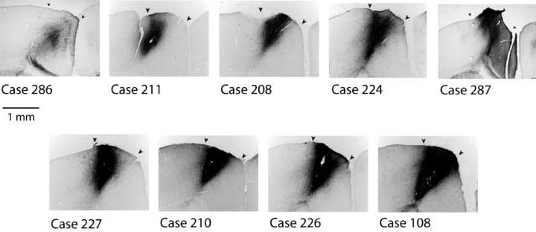
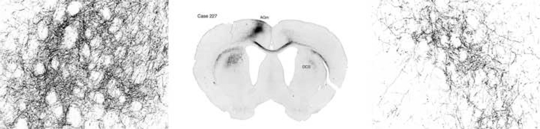
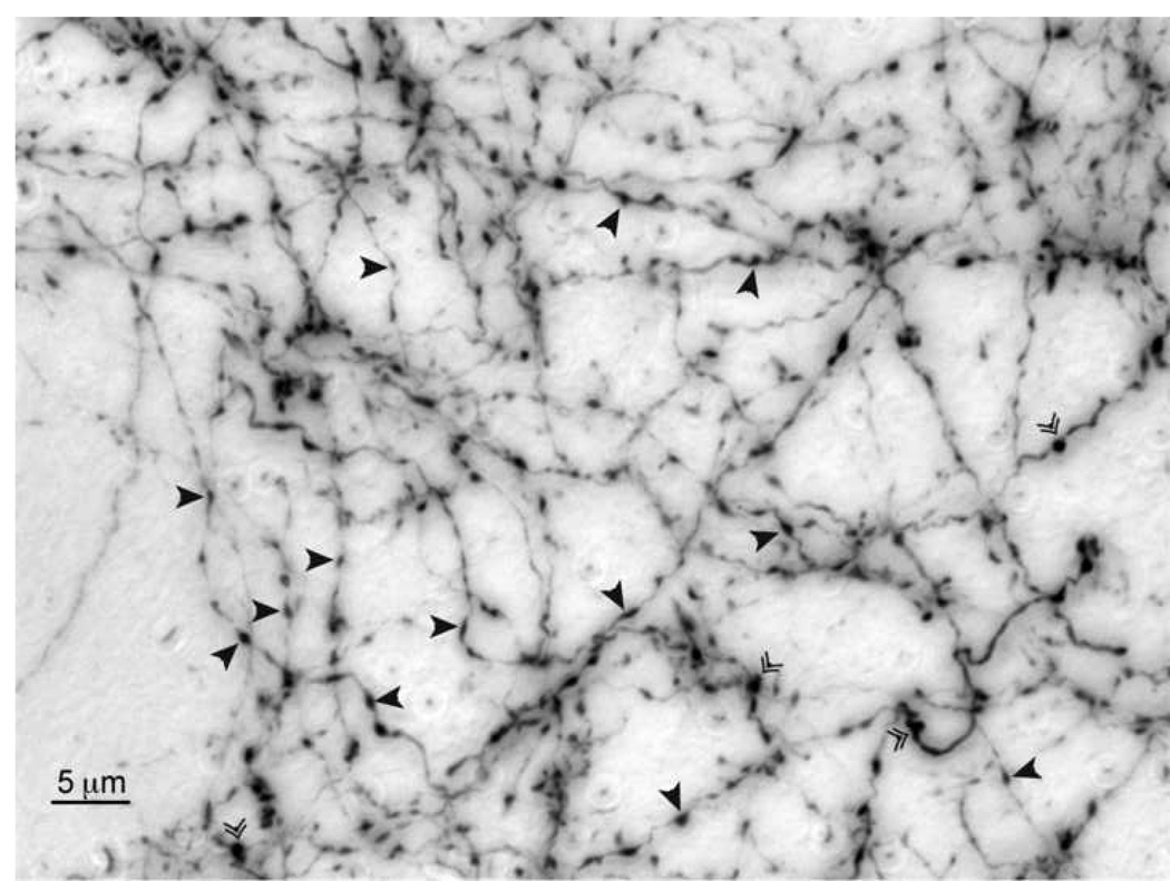
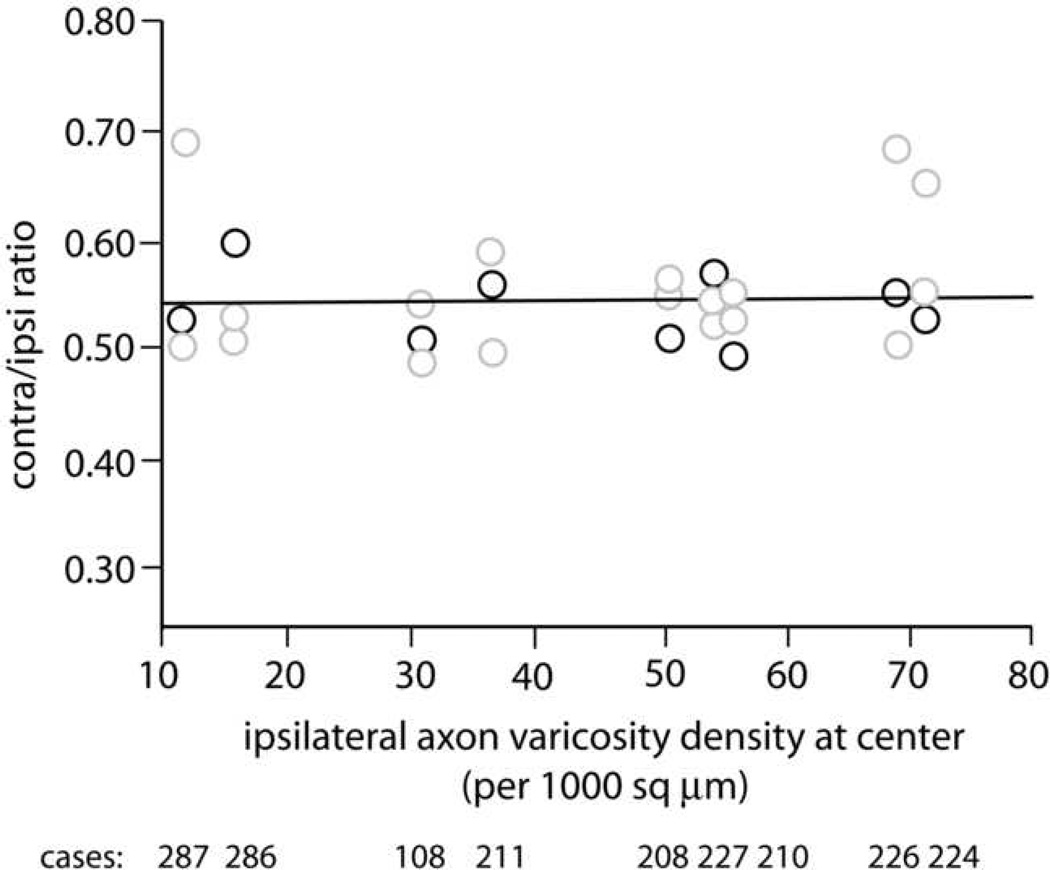
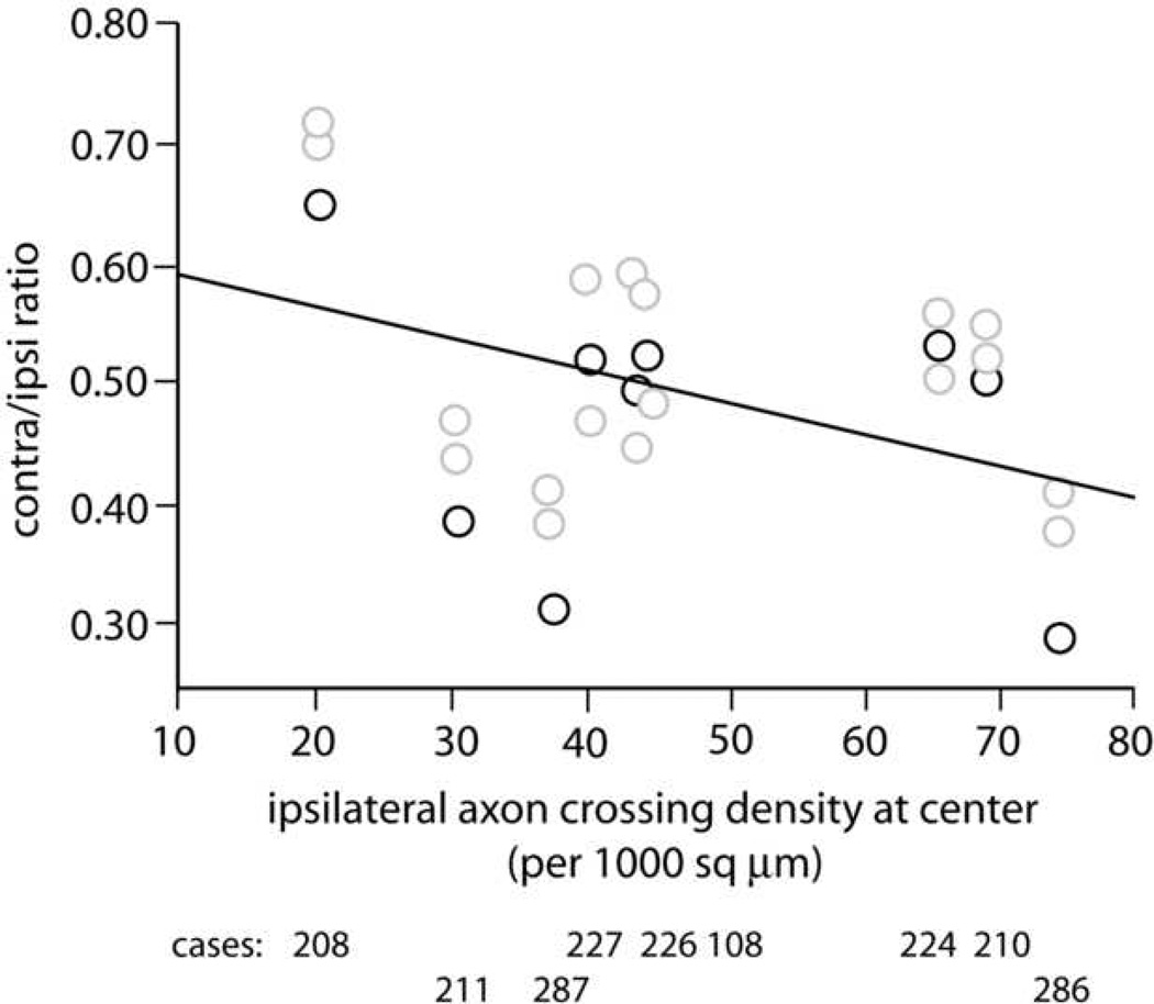
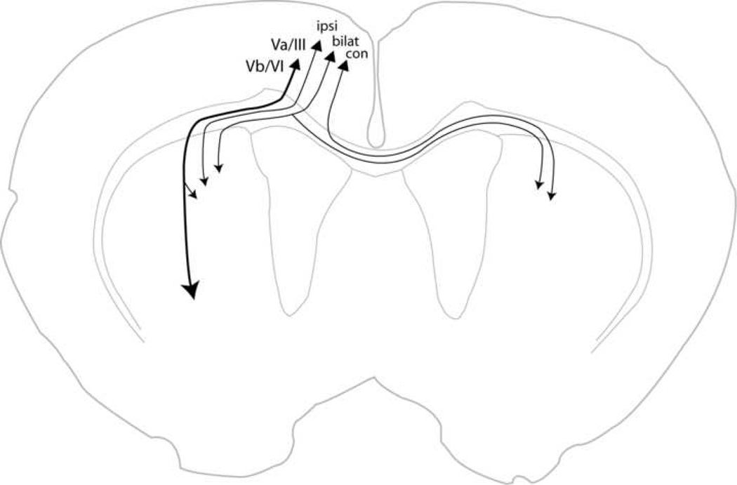
Medial agranular cortex (AGm) has a prominent bilateral projection to the dorsocentral striatum (DCS). We wished to develop a normal baseline by which to assess neuronal plasticity in this corticostriatal system in rats with neglect resulting from a unilateral lesion in AGm, followed by treatment with agents that promote sprouting and functional recovery in other systems. Injections of biotinylated dextran amine were made into AGm in normal rats, and unbiased sampling was used to quantify the density of axons and axonal varicosities present in DCS (the latter represent presynaptic profiles). Labeling density in contralateral DCS is approximately half of that seen in ipsilateral DCS (this ratio is 0.50 for axons, 0.55 for varicosities). The ratio of varicosities is stable over a greater than seven-fold range of absolute densities. There is no consistent relationship between the absolute density of axons and axon varicosities; however, the ratio measures are strongly correlated. We conclude that changes in the contralateral/ipsilateral ratio of axon density after experimental treatments do reflect changes in synaptic density, but axon varicosities are likely to be the most sensitive anatomical parameter by which to assess plasticity at the light microscopic level.


Figures

Figure 1
Figure 1
Nine cases of BDA injection sites in AGm, arranged in order of increasing size and density. The center of each injection site is shown at equivalent magnification and illumination. All injections were confined to AGm and centered at a-p +0.8. Arrowheads indicate boundaries of AGm determined from adjacent Nissl-stained sections.
Figure 2
Figure 2
Axonal labeling in ipsilateral and contralateral dorsocentral striatum in case 227. This representative case illustrates axonal labeling in DCS at low and medium magnification.
Figure 3
Figure 3
Axonal varicosities in dorsocentral striatum. Two size classes of axons are visible. Large caliber axons project to the thalamus, brainstem, and spinal cord. Thinner axons are associated with wholly intratelencephalic projections. Arrowheads denote varicosities. Double arrowheads denote cut axons.
Figure 4
Figure 4
Synaptic density assessed by unbiased sampling of axonal varicosities. Ipsilateral density at the center of the a-p range of labeling was used as an index of the effective size and density of the injection site, and is plotted on the x-axis. Black circles represent contra/ipsi ratios at these central locations, gray circles indicate densities at locations within 240 µm rostral or caudal of the central location. Mean contralateral density is half (0.55) that of the ipsilateral side. The flat slope represents a stable baseline over a greater than seven-fold range of ipsilateral densities. Case numbers are shown below the x-axis, and each is aligned with the data from that brain.
Figure 5
Figure 5
Axon density assessed by unbiased sampling. Similar to the axon varicosity data in Figure 4, ipsilateral axon density at the center of the a-p range of labeling was used as an index of the effective size and density of the injection site, and is plotted on the x-axis. The mean contralateral/ipsilateral ratio of axon density by this measure is 0.50.
Figure 6
Figure 6
Schematic diagram of the four neuron types that contribute to corticostriatal projections. Neurons in layer Vb/VI have large caliber axons that project to thalamus, brainstem, and spinal cord, giving off collaterals in striatum. Neurons in layer Va/III project to ipsilateral striatum, contralateral striatum, or both.

References

    1. Burcham K, Corwin JV, Stoll M, Reep RL. Disconnection of medial agranular and posterior parietal cortex produces multimodal neglect in rats. Behav. Brain Res. 1997;86:41–47. - PubMed
    1. Carman JB, Cowan WM, Powell TPS, Webster KE. A bilateral cortico-striate projection. J. Neurol. Neurosurg. Psychiat. 1965;28:71–77. - PMC - PubMed
    1. Carmichael ST, Chesselet M-F. Synchronous neuronal activity is a signal for axonal sprouting after cortical lesions in the adult. J. Neurosci. 2002;22:6062–6070. - PMC - PubMed
    1. Cheatwood JL, Reep RL, Corwin JV. The associative striatum: cortical and thalamic projections to the dorsocentral striatum. Brain Res. 2003;968:1–14. - PubMed
    1. Chen S, Hillman DE. Robust synaptic plasticity of striatal cells following partial deafferentation. Brain Res. 1990;520:103–114. - PubMed

Publication types