Review
. 2008 Aug;19(4):329-40.
doi: 10.1016/j.semcdb.2008.07.003. Epub 2008 Jul 24.

Paradigm shifts in the cell biology of STAT signaling

Affiliations
Review

Paradigm shifts in the cell biology of STAT signaling

Pravin B Sehgal. Semin Cell Dev Biol. 2008 Aug.

Abstract

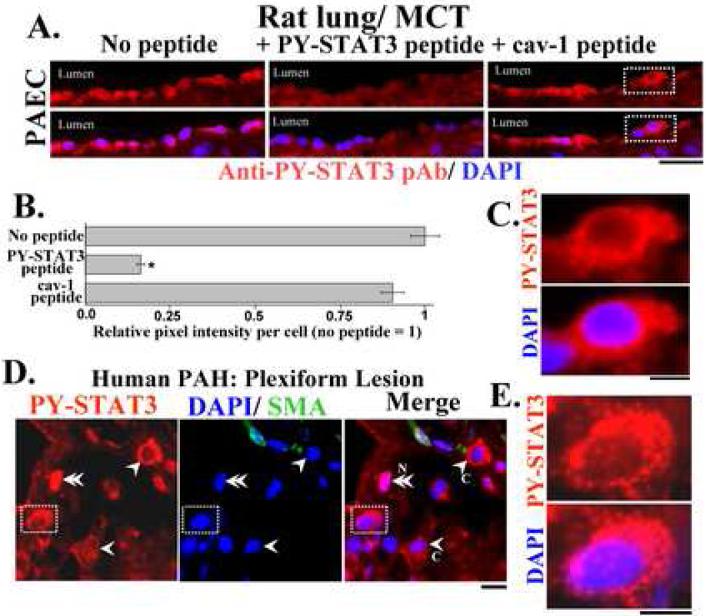
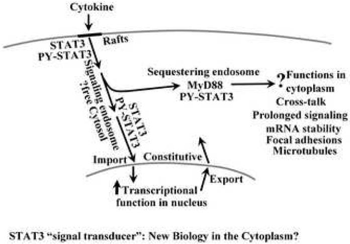
In recent years several of the key tenets of the original cytokine-STAT-signaling paradigm had to be revised. First, the notion that nonphosphorylated "inactive" STATs are present in the cytoplasm as free monomers which dimerized only subsequent to Tyr-phosphorylation has been replaced by the understanding that nonphosphorylated STATs in the cytoplasm exist largely as dimers and high molecular mass "statosome" complexes. Second, the notion that phosphorylation, either of Tyr or Ser residues or both, in STAT species is required for transcriptional activation has been replaced by the realization that nonphosphorylated STATs can be transcriptionally active albeit with respect to sets of target genes distinct from phosphorylated STATs. Third, the notion that it is the activation by phosphorylation of STATs at the plasma membrane that then leads to their import into the nucleus has been replaced by the recognition that even nonphosphorylated STATs shuttle between the cytoplasm and nucleus at all times in a constitutive manner. Fourth, the notion that the trans-cytoplasmic transit of STATs from the plasma membrane to the nuclear import machinery takes place exclusively as a free cytosolic process has been replaced by the understanding that at least a portion of this trans-cytoplasmic transit is mediated via membrane-associated caveolar and endocytic trafficking (the "signaling endosome" hypothesis). Fifth, the targeting and sequestration of activated STAT3 to long-lived endosomes in the cytoplasm requires consideration of STAT3-mediated "signal transduction" from the plasma membrane to cytoplasmic membrane destinations potentially for function(s) in the cytoplasm. Indeed, in tissue sections many discrete histologic cell types display PY-STAT3 almost exclusively in the cytoplasm with little, if any, in the nucleus. New challenges include determining the structural bases for the recruitment of nonphosphorylated dimeric STAT species to the cytosolic face of membranes including at the cytoplasmic tails of respective receptor complexes, the conformational changes subsequent to phosphorylation and the structural bases for the targeting and functions of STAT proteins within the cytoplasm per se.


Figures

Fig. 1
Fig. 1
Superose-6 FPLC analyses of STAT proteins in rat liver cytosol. An aliquot (200 μl) of rat liver cytosol (the 100,000 x g supernatant) was fractionated through Superose-6 FPLC (approximately 1 ml per fraction), and the elution of various STAT proteins was evaluated by SDS-PAGE and Western blotting of 100-μl aliquots of each elutae fraction. Panels A-D show the elution of STAT3, STAT1, STAT5A and STAT5B. Panel E shows the elution of murine anti-STAT3 mAb (IgG) of mass approximately 150-160 kDa off the same column in a separate run. Fr. No., fraction number; void volume of the column is in fractions 7-8. From ref. 7.
Fig. 2
Fig. 2
Immunofluorescence analyses for STAT3 in Hep3B cells in culture showing punctuate cytoplasmic immunostaining and dependence of observations on specific fixation protocol used. Analyses for STAT3 were carried out in untreated and IL-6-treated (10 ng/ml for 30 min) Hep3B cells using the C20 anti-STAT3 rabbit IgG from Santa Cruz, Inc, Santa Cruz, CA using cold methanol-acetone, cold paraformaldehyde-Triton X-100 or formaldehyde (37°C)-Triton X-100 fixation methods. Respective peptide competition controls using relevant and irrelevant (cav-1) peptides were included in the analyses. Scale bar = 25 μm. From ref. 14.
Fig. 3
Fig. 3
Live-cell imaging of the sequestration of fluorescently tagged STAT3 to cytoplasmic sequestering endosomes. Panels A and B. STAT3-GFP (green) transfected Hep3B cells were exposed to IL-6 for 30 min and imaged together with labeling for the lysosomal (LysoTracker in red), endoplasmic reticulum (ER-Tracker in red) or mitochondrial compartments (MitoTracker in blue). Panel C. Sequestration of endogenous native PY-STAT3 in cytoplasmic membrane structures. Replicate Hep3B cells were exposed to IL-6 for 30 min and then sequentially to digitonin (50 μg/ml) in ice-cold 0.25 M sucrose-phosphate-buffered saline (sucrose buffer) and to Brij 58 (0.5% vol/vol in sucrose buffer), fixed with cold paraformaldehyde and immunostained for PY-STAT3. Panels D and E. Cotransfection with an expression construct for the K44A dominant-negative mutant of dynamin II leads to marked accumulation of STAT3 in cytoplasmic vesicles with depletion from the nucleus. All scale bars = 25 μm; adapted from ref. 41.
Fig. 4
Fig. 4
Signal transduction through the cytoplasm: the signaling and sequestering endosome mechanisms. Adapted from ref. 24.
Fig. 5
Fig. 5
Marked cytoplasmic provenance of PY-STAT3 in distinct cell types in lung tissue in pulmonary arterial hypertension assayed in formalin-fixed paraffin-block sections. Panels A and B. Double-label immunofluorescence analyses using anti-PY-STAT3 pAb and DAPI showing PY-STAT3 in the cytoplasm of luminal pulmonary arterial endothelial cells in rats administered monocrotaline 4 wk earlier. Scale bar = 25 μm. Analyses included a peptide competition assay confirming the specificity of the PY-STAT3 pAb used. Per cell integrated pixel intensities were obtained using NIH Image J and expressed in terms of the section stained with PY-STAT3 pAb without any blocking peptide. * P < 0.05 in comparison to the section stained with PY-STAT3 pAb without any peptide, n = 50-60 cells per variable. Panel C. High magnification panels of indicated area in Panel A. Scale bar = 5 μm. Panel D. Plexiform lesion in idiopathic pulmonary arterial hypertension in man showing cells with both cytoplasmic (single arrowheads) and nuclear (double arrowheads) PY-STAT3. Scale bar = 5 μm. Panel E. High magnification panels of indicated area in Panel D. Scale bar = 5 μm. Adapted from ref. 24.
Fig. 6
Fig. 6
Predominant nuclear or cytoplasmic provenance of PY-STAT3 in cells of distinct histologic phenotypes in the human lung. Panel A. Predominantly nuclear PY-STAT3 in cells lining thickened alveolar septa in a patient with pulmonary arterial hypertension. Panel B. Exclusively cytoplasmic PY-STAT3 in the tracheobronchila epithelium in normal lung in cells apparently of the basal cell progenitor phenotype. Panel C. Low and high magnification panels of a peptide competition assays using sequential serial sections of the same block to verify the detection of PY-STAT3 as in panel B. Scale bars = 25 μm except in the low magnification panel in C in which scale bar = 10 μm. (formalin-fixed paraffin-block sections of human lung specimens were kindly provided by Dr. Rubin M. Tuder, Department of Pathology, Johns Hopkins University School of Medicine as in ref. 24).
Fig. 7
Fig. 7
Signal transduction by STAT3 to cytoplasmic destinations.
Fig. 8
Fig. 8
STAT3 and PY-STAT3 during mitosis. Double-label confocal images of cells in mitosis in cultures of Hep3B or of bovine pulmonary arterial endothelial cells were captured au naturel showing the co-localization of STAT3 with clathrin heavy chain (CHC) or with LAMP1 (Hep3B cells) or of PY-STAT3 with CHC (endothelial cells). Scale bar = 10 μm. Adapted from ref. 82.

References

    1. Darnell JE, Kerr IM, Stark GR. Jak-STAT pathways and transcriptional activation in response to IFNs and other extracellular signaling proteins. Science. 1994;264:1415–21. - PubMed
    1. Levy DE, Darnell JE. Stats: transcriptional control and biological impact. Nat Rev Mol Cell Biol. 2002;3:651–62. - PubMed
    1. Mertens C, Darnell JE. Snapshot: JAK-STAT signaling. Cell. 2007;131:612. - PubMed
    1. Sehgal PB, Levy DE, Hirano T, editors. Signal Transducers and Activators of Transcription. Activation and Biology, Kluwer Academic Press; Dordrecht, The Netherlands: 2003. pp. 1–746.
    1. Shuai K, Horvath CM, Huang LH, Qureshi SA, Cowburn D, Darnell JE., Jr Interferon activation of the transcription factor Stat91 involves dimerization through SH-2 phosphotyrosyl peptide interactions. Cell. 1994;76:821–8. - PubMed

Publication types

Substances