. 2008 Sep 5;134(5):877-86.
doi: 10.1016/j.cell.2008.07.041. Epub 2008 Aug 7.

Disease-specific induced pluripotent stem cells

Affiliations

Disease-specific induced pluripotent stem cells

In-Hyun Park et al. Cell. .

Abstract

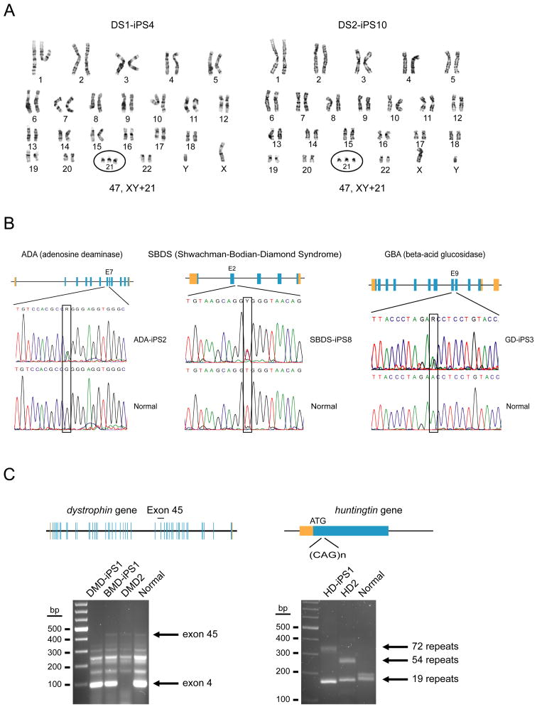
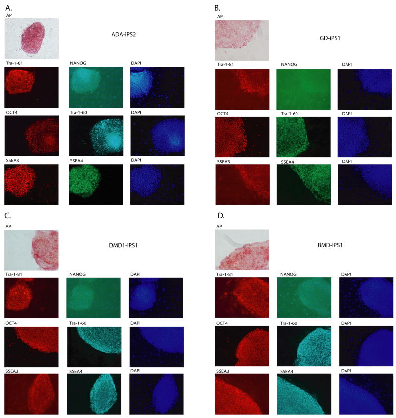
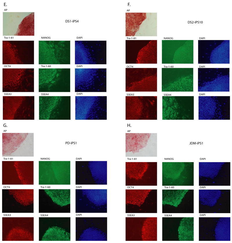
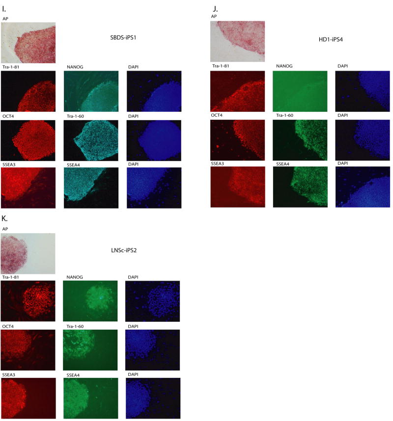
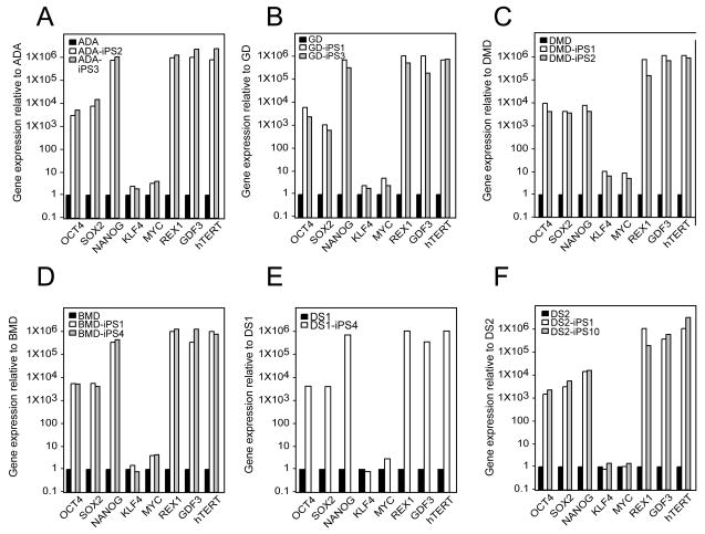
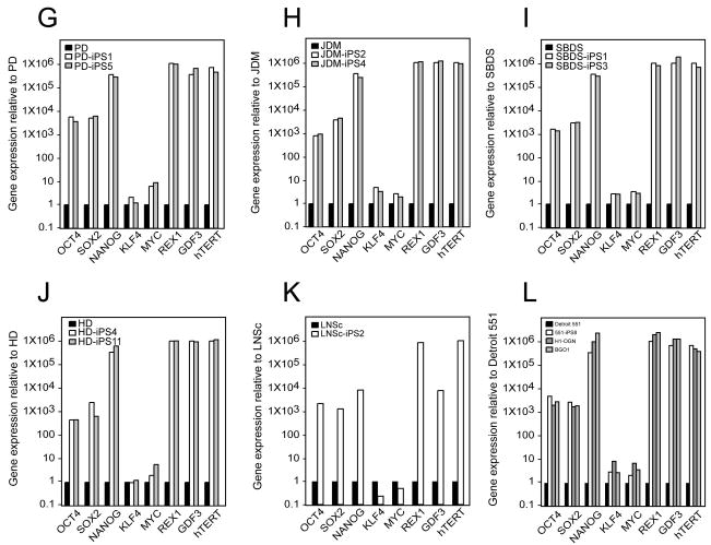
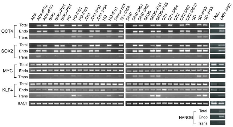
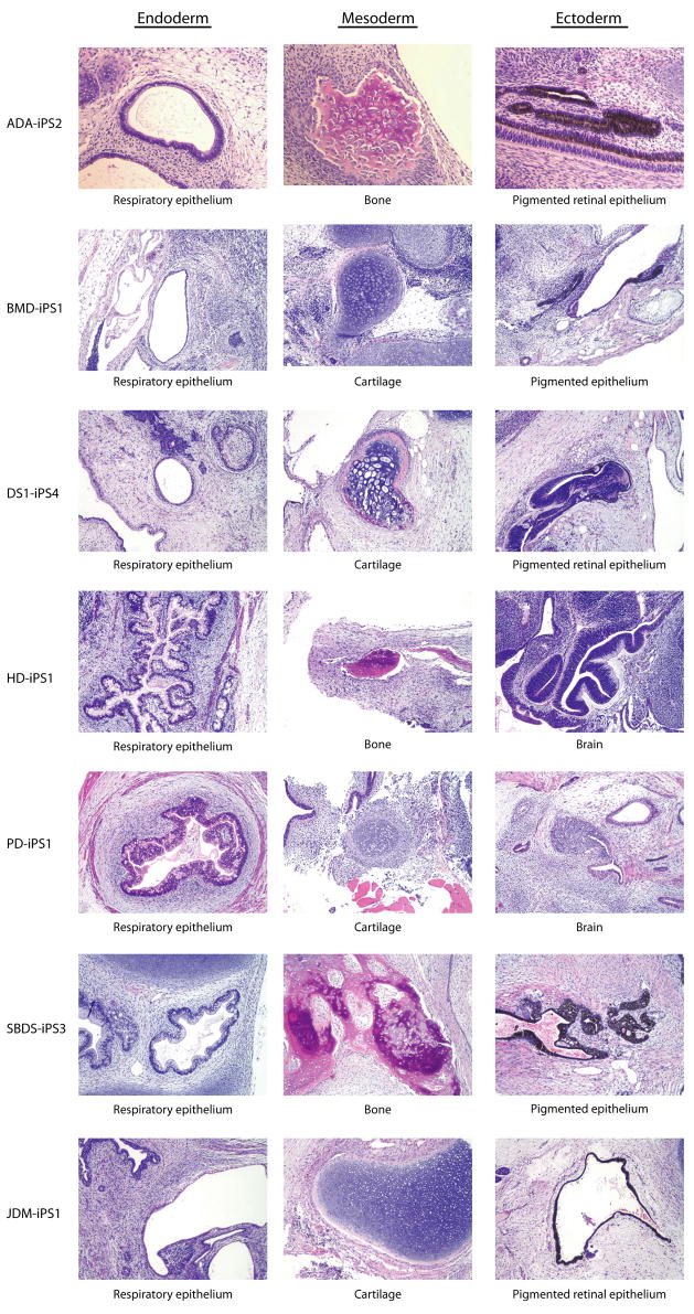
Tissue culture of immortal cell strains from diseased patients is an invaluable resource for medical research but is largely limited to tumor cell lines or transformed derivatives of native tissues. Here we describe the generation of induced pluripotent stem (iPS) cells from patients with a variety of genetic diseases with either Mendelian or complex inheritance; these diseases include adenosine deaminase deficiency-related severe combined immunodeficiency (ADA-SCID), Shwachman-Bodian-Diamond syndrome (SBDS), Gaucher disease (GD) type III, Duchenne (DMD) and Becker muscular dystrophy (BMD), Parkinson disease (PD), Huntington disease (HD), juvenile-onset, type 1 diabetes mellitus (JDM), Down syndrome (DS)/trisomy 21, and the carrier state of Lesch-Nyhan syndrome. Such disease-specific stem cells offer an unprecedented opportunity to recapitulate both normal and pathologic human tissue formation in vitro, thereby enabling disease investigation and drug development.


Figures

Figure 1
Figure 1. Genotypic analysis of disease-specific iPS cell lines
A) Two different, primary fibroblast specimens, DS1 and DS2 from male patients with Down syndrome (trisomy 21) were used to derive DS1-iPS4 and DS2-iPS10. Each has a 47, XY+21 karyotype over several passages (G-banding analysis). B) Fibroblast (ADA and GBA) or bone marrow mesenchymal cells (SBDS) were used to generate iPS lines. Mutated alleles identical to the original specimens were verified by DNA sequencing. Adenosine deaminase deficiency line ADA-iPS2, a compound heterozygote: GGG to GAA double transition in exon 7 of one allele (G216R substitution); the second allele is an exon 10 frame-shift deletion (-GAAGA) (Hirschhorn et al., 1993). Shwachman-Bodian-Diamond syndrome line SBDS-iPS8 is also a compound heterozygote: point mutations at the IV2+2T>C intron 2 splice donor site and an IVS3-1G>A mutation of the SBDS gene (Austin et al., 2005). GD-iPS3 (Gaucher disease type III); a 1226A>G point mutation (N370S substitution) and a guanine insertion at nucleotide 84 of the cDNA (84GG) (Beutler et al., 1991). C) Fibroblasts from patients diagnosed with either Duchenne (DMD) or Becker type muscular dystrophy (BMD): DMD-iPS1 has a deletion over exons 45–52 (multiplex PCR for the dystrophin gene). We could not determine a deletion in BMD-iPS1 using two different multiplex PCR sets though these assays do not cover the entire coding region. DMD2 is a patient control (exon 4 deletion). The control is genomic DNA from a healthy volunteer. Huntington disease (HD) is caused by a tri-nucleotide repeat expansion within the huntingtin locus. DNA sequencing shows that HD-iPS has one normal (<35 repeats) and one expanded allele (72 repeats). HD2 is a positive control from a second Huntington patient with one normal and one expanded allele (54 repeats). The control is genomic DNA from a healthy volunteer.
Figure 2
Figure 2. Patient-derived iPS lines exhibit markers of pluripotency
ADA-iPS2, GD-iPS1, DMD-iPS1, BMD-iPS1, DS1-iPS4, DS2-iPS10, PD-iPS1, JDM-iPS1, SBDS-iPS1, HD-iPS4, LNSc-iPS2, JDM-iPS2 were established from a fibroblast or mesenchymal cells (Table 1). Disease specific iPS cell lines maintain a morphology similar to hES cells when grown in co-culture with mouse embryonic feeder fibroblasts (MEFs). Patient-specific iPS cells express alkaline phosphatase (AP). Also, as shown here via immunohistochemistry, patient-specific cells express pluripotency markers including Tra-1-81, NANOG, OCT4, Tra-1-60, SSEA3 and SSEA4. 4,6-Diamidino-2-phenylindole (DAPI) staining is shown at right and indicates the total cell content per image.
Figure 3
Figure 3. Expression of pluripotency-associate genes is elevated in patient-specific iPS lines relative to their somatic cell controls
In each panel, quantitative real-time PCR (QRT-PCR) assays for OCT4, SOX2, NANOG, REX1, GDF3, and hTERT indicates increased expression in patient-specific iPS cells relative to parent cell lines while expression of KLF4 and cMYC remains largely unchanged. PCR reactions were normalized against internal controls (β-actin) and plotted relative to expression levels in their individual parent fibroblast cell lines. (A) the human iPS lines ADA-iPS2 and –iPS3 are derived from the adenosine deaminase deficiency-severe combined immunodeficiency fibroblast line ADA. (B) GD-iPS1 and –iPS3 are derived from the Gaucher disease type III fibroblast line GD. (C) DMD-iPS1 and –iPS2 are derived from the Duchenne muscular dystrophy fibroblast line DMD. (D) BMD-iPS1 and –iPS4 are derived from the Becker muscular dystrophy line BMD. (E) DS1-iPS4 is derived from the Down syndrome fibroblast line DS1. (F) DS2-iPS1 and –iPS10 are derived from the Down syndrome fibroblast line DS2. (G) PD-iPS1 and –iPS5 are derived from the Parkinson disease fibroblast line PD. (H) JDM-iPS2 and –iPS4 are derived from the juvenile-onset, type 1 diabetes mellitus line JDM. (I) SBDS-iPS1 and –iPS3 are derived from the Shwachman-Bodian-Diamond syndrome bone marrow mesenchymal fibroblast line SBDS. (J) HD-iPS4 and –iPS11 are derived from the Huntington disease fibroblast line HD. (K) LNSc-iPS1 and –iPS2 are derived from the Lesch-Nyhan syndrome carrier fibroblast line LNSc. (L) Detroit 551 human fibroblasts are used as the standard here in order to demonstrate the previously described expression pattern in Detroit 551 derived iPS cells (551-iPS8) relative to two bona fide hES cell lines: H1-OGN and BG01.
Figure 4
Figure 4. Pluripotency-promoting genes are chiefly expressed from the endogenous loci in patient-specific iPS lines, while the virally-delivered transgene is predominantly silenced
The patient-specific iPS cell lines shown here are preceded by their parental fibroblast controls (from left to right at top): adenosine deaminase deficiency-associate severe combined immunodeficiency (ADA), Becker muscular dystrophy (BMD), Parkinson disease (PD), juvenile type one diabetes mellitus (JDM), Huntington disease (HD), Detroit 551 control cells, Duchenne muscular dystrophy (DMD), Shwachman-Bodian-Diamond syndrome (SBDS), Down syndrome (DS), Gaucher disease type III (GD), and Lesch-Nyhan syndrome carrier (LNSc). The semi-quantitative expression (RT-PCR) of the four pluripotency-promoting genes used in the reprogramming process, OCT4, SOX2, cMYC, KLF4 and NANOG is shown for each line using amplification conditions specific to the endogenous (Endo) or virally-delivered transgene (Trans) as well as the total expression for each (Total). Beta-actin is shown at the bottom as a loading control for each lane.
Figure 5
Figure 5. Differentiation of patient-specific iPS lines reveals lineage-specific gene expression and mature cell formation
A) At top (from left to right) are nine iPS cell lines in their undifferentiated (U) or differentiated (D) state. The lines are: adenosine deaminase deficiency-associated severe combined immunodeficiency (ADA), juvenile-onset type one diabetes mellitus (JDM), Down syndrome 1 (DS1), Gaucher disease type III (GD), Huntington disease (HD), Duchenne muscular dystrophy (DMD), Down syndrome 2 (DS2), and normal control Detroit 551 (551) cells. Differentiation (D) of these patient-specific iPS cells as embryoid bodies (EB) followed by RT-PCR analysis shows upregulated expression of lineage markers from the three embryonic germ layers relative to their undifferentiated controls (U) including: GATA4 and AFP (endoderm), RUNX1 and Brachyury (mesoderm), and Nestin and NCAM (ectoderm). Beta-actin serves as a positive amplification control for each. B) Differentiation of ADA-iPS2, a representative patient-specific iPS cell line, as embryoid bodies (EB) is highly reminiscent of that using hES cells where tight clusters of differentiating cells are well-formed by day 7 which will cavitate, becoming cystic, by day 10. Hematopoietic differentiation of patient-specific iPS cells yields various blood cell types in semi-solid methylcellulose colony-forming assays including burst-forming unit-erythroid (BFU-E) which are derivative of red blood cell progenitor cells.
Figure 6
Figure 6. Patient-specific iPS lines form teratomas in immunodeficient mice
Shown here are the representative series of hematoxylin-eosin (H/E) stained sections from a formalin fixed teratoma produced from ADA-iPS2, BMD-iPS1, DS1-iPS4, HD-iPS1, PD-iPS1, SBDS-iPS3, and JDM-iPS1 cell lines. They formed mature, cystic teratomas with tissues representing all three embryonic germ layers including: respiratory epithelium (endoderm), bone and cartilage (mesoderm), and pigmented retinal epithelium and immature neural tissue (ectoderm).

References

    1. Antonarakis SE, Lyle R, Dermitzakis ET, Reymond A, Deutsch S. Chromosome 21 and down syndrome: from genomics to pathophysiology. Nat Rev Genet. 2004;5:725–738. - PubMed
    1. Austin KM, Leary RJ, Shimamura A. The Shwachman-Diamond SBDS protein localizes to the nucleolus. Blood. 2005;106:1253–1258. - PMC - PubMed
    1. Beggs AH, Koenig M, Boyce FM, Kunkel LM. Detection of 98% of DMD/BMD gene deletions by polymerase chain reaction. Hum Genet. 1990;86:45–48. - PubMed
    1. Beutler E, Gelbart T, Kuhl W, Sorge J, West C. Identification of the second common Jewish Gaucher disease mutation makes possible population-based screening for the heterozygous state. Proc Natl Acad Sci U S A. 1991;88:10544–10547. - PMC - PubMed
    1. Bittles AH, Bower C, Hussain R, Glasson EJ. The four ages of Down syndrome. Eur J Public Health. 2007;17:221–225. - PubMed

Publication types