. 2008 Aug 8;31(3):383-94.
doi: 10.1016/j.molcel.2008.06.018.

General and versatile autoinhibition of PLC isozymes

Affiliations

General and versatile autoinhibition of PLC isozymes

Stephanie N Hicks et al. Mol Cell. .

Abstract

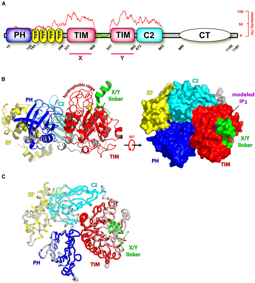
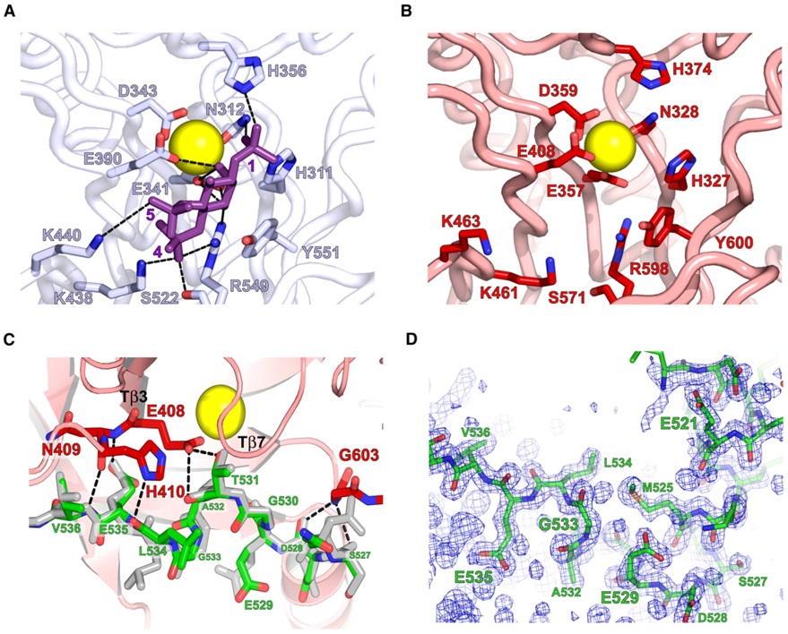
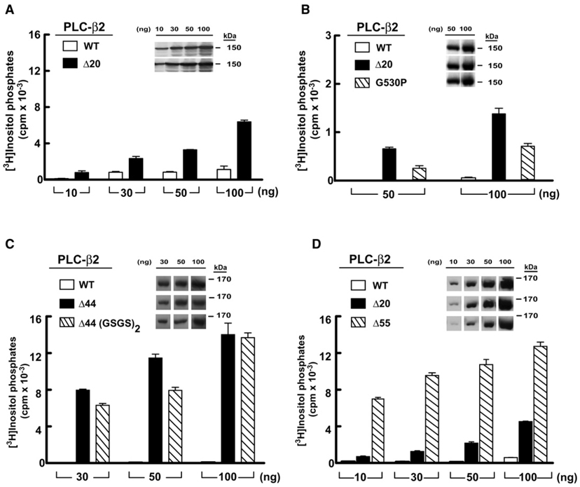
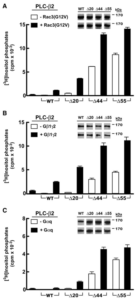
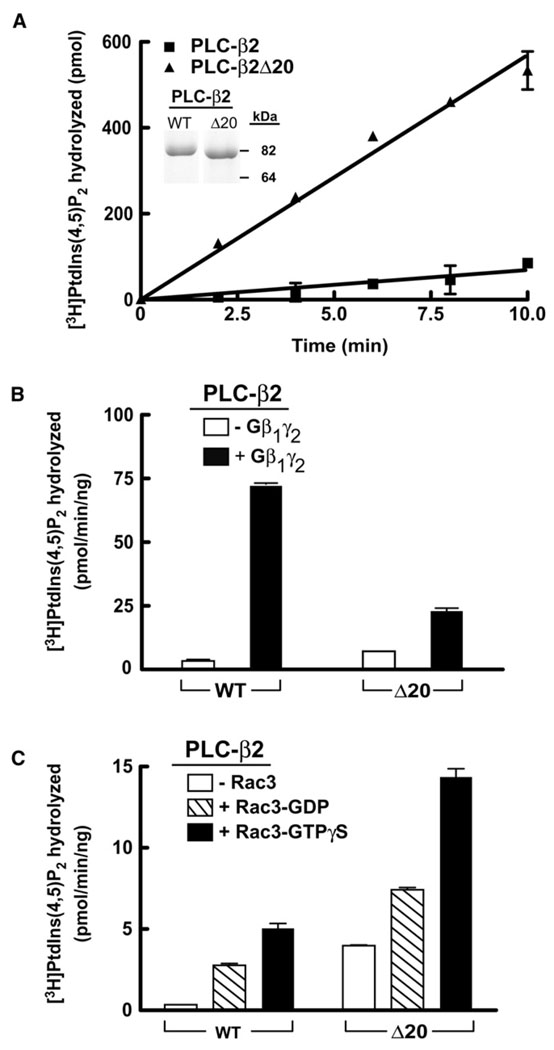
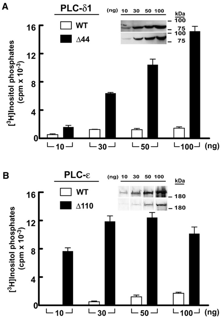
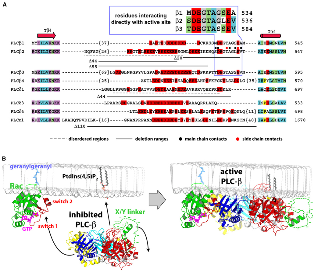
Phospholipase C (PLC) isozymes are directly activated by heterotrimeric G proteins and Ras-like GTPases to hydrolyze phosphatidylinositol 4,5-bisphosphate into the second messengers diacylglycerol and inositol 1,4,5-trisphosphate. Although PLCs play central roles in myriad signaling cascades, the molecular details of their activation remain poorly understood. As described here, the crystal structure of PLC-beta2 illustrates occlusion of the active site by a loop separating the two halves of the catalytic TIM barrel. Removal of this insertion constitutively activates PLC-beta2 without ablating its capacity to be further stimulated by classical G protein modulators. Similar regulation occurs in other PLC members, and a general mechanism of interfacial activation at membranes is presented that provides a unifying framework for PLC activation by diverse stimuli.


Figures

Figure 1
Figure 1. Overall Structure of PLC-β2
(A) PLC-β isozymes are composed of an N-terminal pleckstrin homology (PH) domain, an array of EF hands, a catalytic TIM barrel split by a highly degenerate linker sequence (green), a C2 domain, and a C-terminal coiled-coil (CT) domain necessary for homodimerization. Sequence conservation of all human PLCs is graphed (red trace) relative to their shared domain architecture, X and Y regions of high sequence conservation are indicated, and absolute domain borders are listed for human PLC-β2. (B) The X/Y linker occludes the active site of PLC-β2. The 1.6 Å resolution structure of PLC-β2 is depicted in ribbon form (left panel) with domain boundaries colored as in (A) and the approximate membrane-binding surface at the top. Also shown are the calcium cofactor (yellow sphere) within the active site and the hydrophobic ridge that is a major point of contact with membranes. Surface representation of PLC-β2 (right panel) rotated 90° with respect to the left panel emphasizes the occlusion of the active site within the TIM barrel by the X/Y linker. For reference, superposition of the active site of PLC-δ1 containing IP3 and PLC-β2 was used to dock IP3 (purple) into the TIM barrel of PLC-β2. (C) Superimposition of the crystal structures of the isolated PLC-β2 fragment colored as in (B) and the equivalent fragment from the Rac1-bound form (gray; PDB ID code 2FJU).
Figure 2
Figure 2. Structural Details of the Active Site of PLC-β2
(A and B) Active site residues in PLC-δ1 that coordinate calcium (yellow) and interact (dashed lines) with IP3 (purple) are conserved in (B) PLC-β2. (C) An extended portion of the X/Y linker (green; residues 527–536) participates in a set of H bonds (dashed lines) with active site residues (red) of PLC-β2. The equivalent portion of the linker from the structure of PLC-β2 bound to Rac1 is shown in gray. (D) Simulated annealing omit map (2Fo – Fc; contoured at 1σ) highlighting the density for residues 517–537 of the X/Y linker.
Figure 3
Figure 3. Deletion of the X/Y Linker Constitutively Activates PLC-β2
COS-7 cells were transfected with the indicated amounts of DNA encoding either wild-type (white bars) or the indicated mutant forms of PLC-β2 (black or hatched bars). (A) PLC-β2Δ20 lacking the ordered part of the X/Y linker. (B) PLC-β2 harboring a G530P substitution within the X/Y linker. (C) PLC-β2Δ44 lacking the majority of the disordered portion of the linker. (D) PLC-β2Δ55 lacking the linker with the exception of residues that occupy the active site. [3H]Inositol phosphate accumulation in cells transfected with vector alone was subtracted from all measurements. Data shown are the mean ± SD of triplicate samples and are representative of data obtained in three or more experiments. Insets confirm equivalent expression of PLC-β isozymes by western blotting of COS-7 cell lysate.
Figure 4
Figure 4. Activation of PLC-β2 by G Protein Activators Is Retained in PLC-β2 Lacking the Majority of the X/Y Linker
COS-7 cells were transfected with DNA encoding either PLC-β2 or the indicated PLC-β2 deletion forms in the presence or absence of 30 ng of constitutively active Rac3(G12V) (A), 150 ng of each subunit of Gβ1γ2 (B), or 100 ng of Gαq (C) prior to quantification of [3H]inositol phosphates and vector subtraction. All transfections used 30 ng of each PLC form except for (C), where 10 ng of each PLC was transfected. Endogenous activity resulting from transfection of either G protein or Rac3(G12V) alone is indicated by the first bar on each plot. Data are the mean ± SD of triplicate samples and are representative of results from three or more experiments. PLC-β2 expression was assessed by western blotting (insets).
Figure 5
Figure 5. Purified PLC-β2 Lacking the Ordered Region of the X/Y Linker Is Constitutively Active and Is Further Stimulated by Gβ1γ2 and Rac1 upon Reconstitution in Lipid Vesicles
(A) Purified PLC-β2 (5 ng) or PLC-β2Δ20 (5 ng) was incubated with mixed detergent- lipid vesicles containing [3H]PtdIns(4,5)P2 for the indicated times at 30°C. Purity of proteins (2 µg, inset) was assessed by SDS-PAGE followed by staining with Coomassie Brilliant Blue. Hydrolysis of [3H]PtdIns(4,5)P2 was quantified in phospholipid vesicles reconstituted with (B) purified Gβ1γ2 (250 nM, final concentration) or (C) purified Rac1 (300 nM, final concentration) preincubated with 10 µM GDP or GTPγS for 30 min. [3H]PtdIns(4,5)P2 hydrolysis was initiated by addition of either 1 ng or 5 ng PLC-β2 or PLC-β2Δ20, and incubations were terminated after 10 min at 30°C. The data are the mean ± range of duplicate determinations, and the results are representative of three individual experiments.
Figure 6
Figure 6. Deletion of the X/Y Linker Constitutively Activates Diverse PLC Isozymes
COS-7 cells were transfected with PLC-δ1 (A) or PLC-ε (B) in tandem with the respective forms of these isozymes lacking the X/Y linker. In all cases, accumulation of inositol phosphates in cells transfected with vector alone was subtracted. Data are the mean ± SD for triplicate samples, and the results are representative of at least three independent experiments. Western blotting (insets) confirms expression of PLC isozymes in COS-7 cell lysate.
Figure 7
Figure 7. Sequence Details of the X/Y Linker within PLC Isozymes and Model for Interfacial Activation of PLC-β2
(A) The majority of human PLCs possess a region of dense negative charge (red boxes) within their X/Y linkers. Otherwise, these insertions are highly variable in length, possess no significant sequence conservation, and form loops between highly conserved secondary structure elements (Tβ4, Tα4) of the TIM barrel. Number of residues not listed are in brackets; disordered regions within the crystal structures of PLC-δ1 and PLC-β2 are underlined with dashed lines (gray); and regions deleted for biochemical analyses are underlined (solid black). Also highlighted are residues within the X/Y linker of PLC-β2 that participate in direct main-chain (black circles) and side-chain (red circles) H-bonds with the TIM barrel. This region is expanded above the alignment for the PLC-β isozymes. (B) Basally inactive PLC-β2 is cytosolic and inhibited by occlusion of its active site by the ordered portion (green) and the disordered portion (dotted green line with associated negative charge represented by dashes) of the X/Y linker (left panel). Membrane-resident GTP-bound Rac (green with switch regions red) recruits and orients PLC-β2 at membranes through interaction of the switch regions with the PH domain (blue) of PLC-β2. Upon engagement of PLC-β2 by Rac, the X/Y linker region is repelled from negatively charged membranes, which frees the active site to extract partially a molecule of PtdIns(4,5)P2 from the membrane in preparation for its hydrolysis into IP3 and DAG. X/Y linkers within other PLCs are envisioned to behave similarly. Arrows indicate movement of PLC-β2 and the X/Y linker.

References

    1. Barr AJ, Ali H, Haribabu B, Snyderman R, Smrcka AV. Identification of a region at the N-terminus of phospholipase C-β3 that interacts with G protein βγ subunits. Biochemistry. 2000;39:1800–1806. - PubMed
    1. Blank JL, Shaw K, Ross AH, Exton JH. Purification of a 110-kDa phosphoinositide phospholipase C that is activated by G-protein βγsubunits. J. Biol. Chem. 1993;268:25184–25191. - PubMed
    1. Boyer JL, Waldo GL, Harden TK. βγ-subunit activation of G-protein- regulated phospholipase C. J. Biol. Chem. 1992;267:25451–25456. - PubMed
    1. Brunger AT, Adams PD, Clore GM, DeLano WL, Gros P, Grosse-Kunstleve RW, Jiang JS, Kuszewski J, Nilges M, Pannu NS, et al. Crystallography & NMR system: a new software suite for macromolecular structure determination. Acta Crystallogr. D Biol. Crystallogr. 1998;54:905–921. - PubMed
    1. (CCP4) Collaborative Computational Project Number 4. The CCP4 suite: programs for protein crystallography. Acta. Cryst. D. 1994;50:760–763. - PubMed

Publication types

MeSH terms

Associated data