. 2008 Aug 8;31(3):415-21.
doi: 10.1016/j.molcel.2008.06.008.

The paracaspase MALT1 controls caspase-8 activation during lymphocyte proliferation

Affiliations

The paracaspase MALT1 controls caspase-8 activation during lymphocyte proliferation

Holli Kawadler et al. Mol Cell. .

Abstract

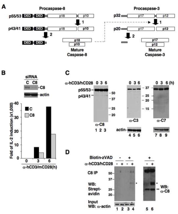
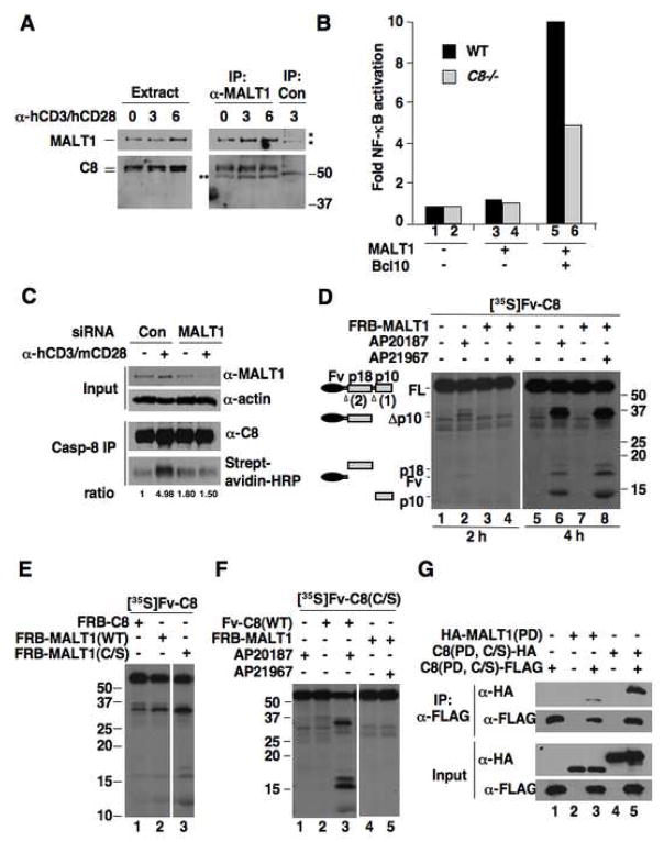
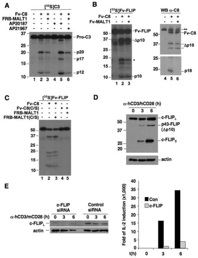
Caspase-8, an initiator caspase involved in lymphocyte apoptosis, is paradoxically required for lymphocyte proliferation. It is not understood how caspase-8 is controlled during antigenic signaling to allow for activation while averting the triggering of apoptosis. Here, we show that caspase-8 undergoes limited activation upon antigenic stimulation, and this activation is dependent on the paracaspase MALT1. The paracaspase domain of MALT1, in a protease-independent manner, induces caspase-8 activation through direct association. MALT1 diminishes the activation of apoptotic effector caspases, but it does not alter the activity of caspase-8 toward c-FLIP(L), which is required for antigenic signaling. Mutants of MALT1 that fail to activate caspase-8 and permit c-FLIP(L) cleavage cannot facilitate NF-kappaB activation or IL-2 induction. Our results reveal a mechanism that utilizes a protease potentially deadly to the cell for proliferative signaling and demonstrate a functional connection between the caspase and paracaspase families to enable nonapoptotic processes.


Conflict of interest statement

The authors declare no conflict of interest.

Figures

Figure 1
Figure 1. Proliferative function of caspase-8 is related to its activation but not full processing
(A) Schematic of caspase-8 and caspase-3 processing. (B) Human primary CD4+ T cells transfected with control (C) or caspase-8 (C8) siRNA plus m/hCD28 were stimulated with α-hCD3/mCD28 antibodies. Caspase-8 expression was analyzed by immunoblotting (top), and IL-2 induction by quantitative RT-PCR (bottom). (C) CD4+ T cells were stimulated with α-hCD3/hCD28 for the indicated durations, and processing of caspase-8, caspase-3 (C3), and caspase-7 (C7) was analyzed by immunoblotting. (D) CD4+ cells treated with α-hCD3/hCD28 were incubated with biotin-zVAD-fmk or DMSO. Lysates were immunoprecipitated with α-caspase-8 and analyzed with streptavidin-HRP (left) or α-caspase-8 (right). * uncharacterized bands. ** IgG light chain bands.
Figure 2
Figure 2. MALT1 associates with caspase-8 and promotes caspase-8 activation through the paracaspase domain
(A) CD4+ cells were treated with α-hCD3/hCD28 or left untreated. Lysates were immunoprecipitated with α-MALT1 or control antibody, followed by Western blot analysis. *, nonspecific bands. **, IgG heavy chain bands. (B) Wild type or caspase-8 deficient Jurkat cells were transfected with MALT1 and Bcl10 as indicated, together with an NF-κB-responsive firefly luciferase reporter plasmid. Fold activation of NF-κB are relative to vector-transfected wild type Jurkat cells and are normalized to a co-transfected Renilla luciferase control. (C) CD4+ cells transfected with control or MALT1 siRNA plus m/hCD28 were stimulated with α-hCD3/mCD28 in the presence of biotin-zVAD-fmk or DMSO. Lysates were immunoprecipitated with α-caspase-8. Lysates and immunoprecipitates were analyzed by Western blot. The relative ratios of biotin-zVAD-labeled caspase-8 to total caspase-8 are given. (D) In vitro-translated, 35S-labeled Fv-caspase-8 was treated with the homo-dimerizer AP20187 or co-incubated with non-isotope-labeled FRB-MALT1 and treated with the hetero-dimerizer AP21967. 35S-labeled proteins were visualized by autoradiography. See Supp. Fig. 2 for relative expression of unlabeled proteins. (E) [35S]Fv-caspase-8 was incubated with FRB-C8 or -MALT1 fusion proteins in the presence of AP21967. The two parts are from the same exposure of the same blot. (F) [35S]Fv-caspase-8(C/S) was incubated with Fv-C8 or FRB-MALT1 and dimerizers. The two parts are from the same exposure of the same blot. (G) The tagged protease domains (PD) of caspase-8 and MALT1 were expressed in 293T cells as indicated and their interactions were detected by co-immunoprecipitation assay.
Figure 3
Figure 3. The MALT1-activated caspase-8 cannot generate the apoptotic form of caspase-3 but is capable of cleaving c-FLIPL
(A) [35S]Caspase-3 was incubated with Fv-Caspase-8 alone or Fv-Caspase-8 plus FRB-MALT1 with or without dimerizer. (B) [35S]Fv-FLIP was incubated with Fv-C8 and Fv-MALT1 as indicated. The reaction mixes were analyzed by autoradiography (left) and anti-caspase-8 immunoblotting (right). *, the large subunit of FLIP that is generated in vitro. **, endogenous caspase-8 in the reticulocyte lysates. (C) [35S]Fv-FLIP was co-incubated with the indicated Fv-C8 and FRB-MALT1 proteins. The reaction mixes were analyzed as in (A). (D) CD4+ T cells were stimulated with α-hCD3/hCD28. c-FLIP proteins and actin were detected by Western blot. (E) Human CD4+ T cells were transfected with control or c-FLIP siRNA plus m/hCD28. Cells were stimulated with α-hCD3/mCD28 beads. IL-2 induction was measured by quantitative RT-PCR.
Figure 4
Figure 4. MALT1-induced caspase-8 activation is required for TCR signaling
(A) Schematic representation of MALT1 mutations. (B) [35S]Fv-C8 was incubated with FRB fusion of caspase-8, MALT1 and MALT1 mutants in the presence of AP21967. See Supp. Figure 2 for relative expression of unlabeled proteins. (C) [35S]Fv-FLIP was incubated with equal amounts of Fv-C8 and Fv-MALT1 proteins as indicated. (D) Jurkat cells were transfected with MALT1 proteins with or without Bcl10, plus an NF-κB responsive luciferase reporter plasmids. Data is representative of three independent experiments. (E) Jurkat cells were transfected with MALT1 plasmids with or without Bcl10. IL-2 mRNA expression was measured by quantitative RT-PCR. Data is representative of three independent experiments. (F) Differential activation of caspase-8 in proliferative and apoptotic signaling. Whereas caspase-8 is activated by homo-dimerization during apoptotic signaling, caspase-8 hetero-dimerizes with MALT1 in response to TCR signaling. This hetero-dimer lacks the capacity to activate apoptotic substrates, but retains activity towards c-FLIPL to enable proliferation.

References

    1. Boatright KM, Renatus M, Scott FL, Sperandio S, Shin H, Pedersen IM, Ricci JE, Edris WA, Sutherlin DP, Green DR, Salvesen GS. A unified model for apical caspase activation. Mol Cell. 2003;11:529–541. - PubMed
    1. Chang DW, Xing Z, Capacio VL, Peter ME, Yang X. Interdimer processing mechanism of procaspase-8 activation. Embo J. 2003;22:4132–4142. - PMC - PubMed
    1. Chang DW, Xing Z, Pan Y, Algeciras-Schimnich A, Barnhart BC, Yaish-Ohad S, Peter ME, Yang X. c-FLIP(L) is a dual function regulator for caspase-8 activation and CD95-mediated apoptosis. Embo J. 2002;21:3704–3714. - PMC - PubMed
    1. Chang DW, Yang X. Activation of procaspases by FK506 binding protein-mediated oligomerization. Sci STKE. 2003;2003:PL1. - PubMed
    1. Chau H, Wong V, Chen NJ, Huang HL, Lin WJ, Mirtsos C, Elford AR, Bonnard M, Wakeham A, You-Ten AI, et al. Cellular FLICE-inhibitory protein is required for T cell survival and cycling. J Exp Med. 2005;202:405–413. - PMC - PubMed

Publication types

MeSH terms

Substances