. 2008 Oct 1;322(1):179-89.
doi: 10.1016/j.ydbio.2008.07.022. Epub 2008 Jul 26.

GATA4 mediates gene repression in the mature mouse small intestine through interactions with friend of GATA (FOG) cofactors

Affiliations

GATA4 mediates gene repression in the mature mouse small intestine through interactions with friend of GATA (FOG) cofactors

Eva Beuling et al. Dev Biol. .

Abstract

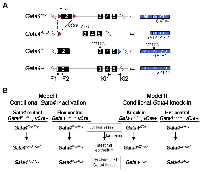
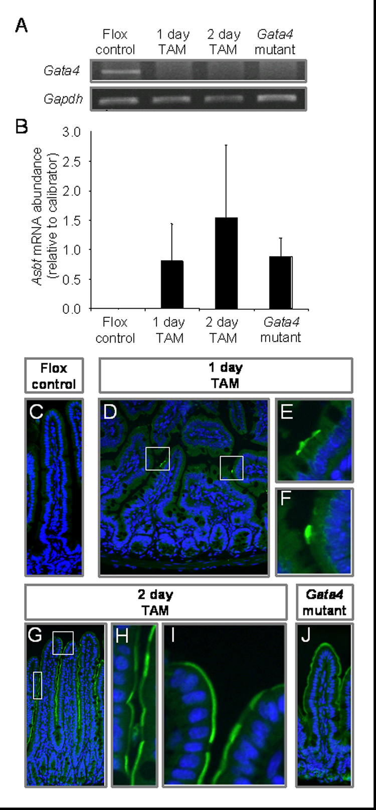
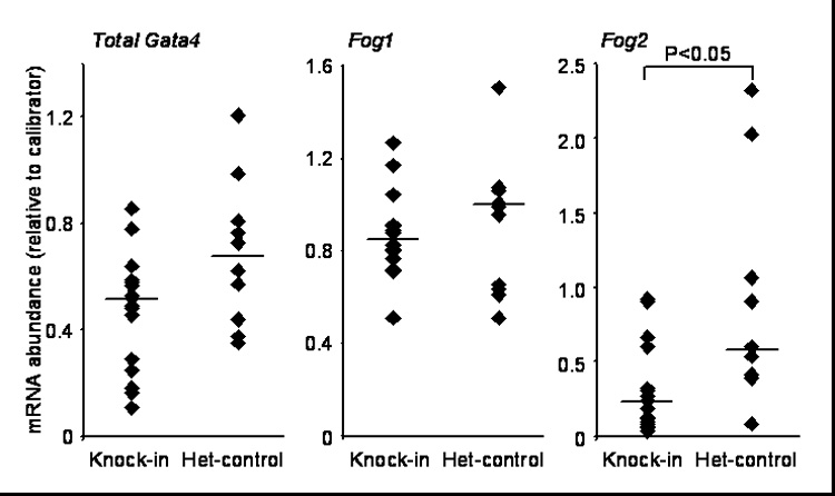
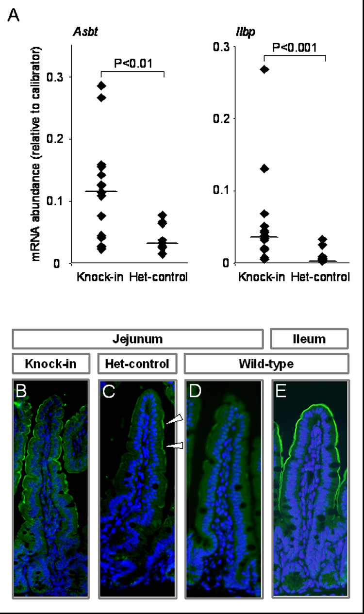
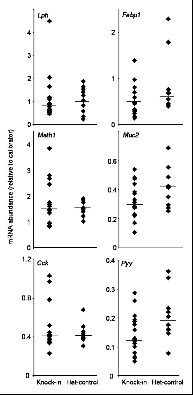
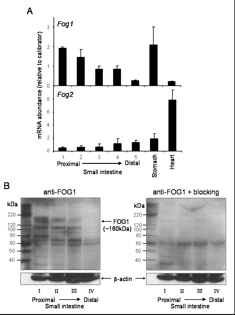
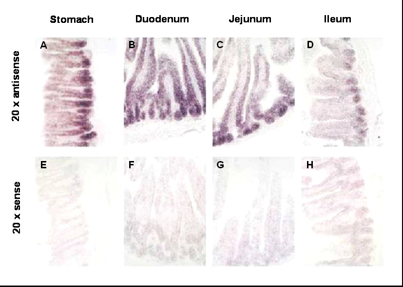
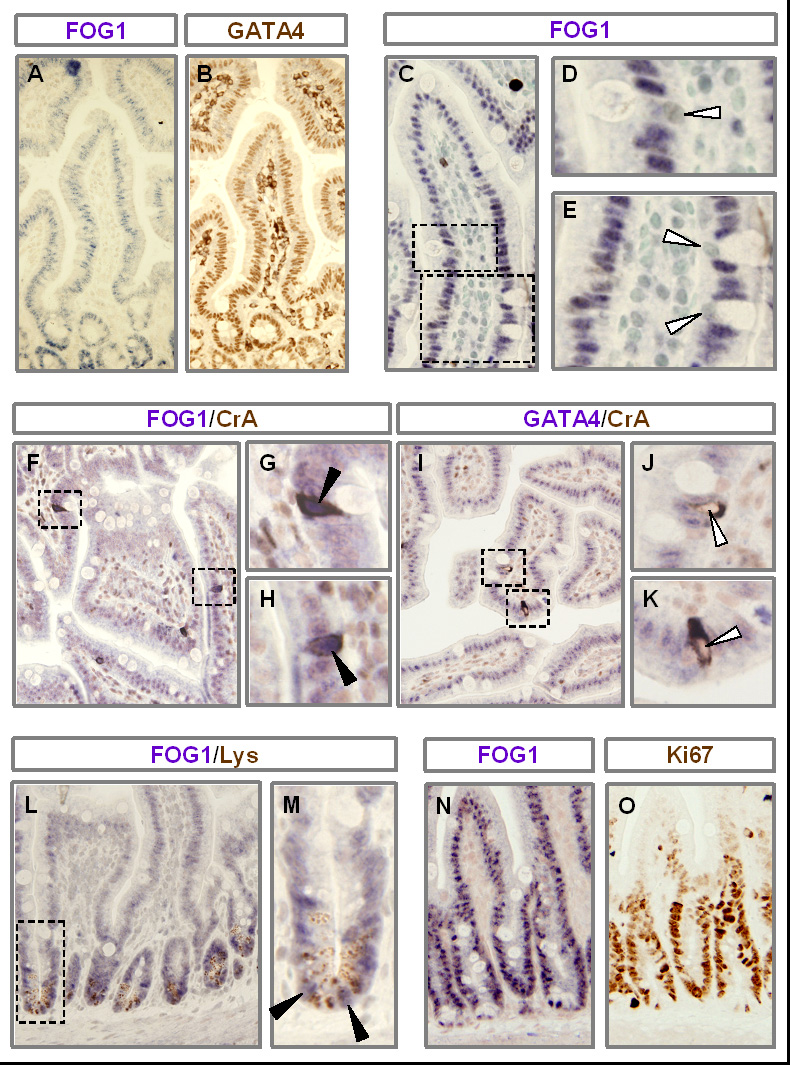
GATA4, a transcription factor expressed in the proximal small intestine but not in the distal ileum, maintains proximal-distal distinctions by multiple processes involving gene repression, gene activation, and cell fate determination. Friend of GATA (FOG) is an evolutionarily conserved family of cofactors whose members physically associate with GATA factors and mediate GATA-regulated repression in multiple tissues. Using a novel, inducible, intestine-specific Gata4 knock-in model in mice, in which wild-type GATA4 is specifically inactivated in the small intestine, but a GATA4 mutant that does not bind FOG cofactors (GATA4ki) continues to be expressed, we found that ileal-specific genes were significantly induced in the proximal small intestine (P<0.01); in contrast, genes restricted to proximal small intestine and cell lineage markers were unaffected, indicating that GATA4-FOG interactions contribute specifically to the repression function of GATA4 within this organ. Fog1 mRNA displayed a proximal-distal pattern that parallels that of Gata4, and FOG1 protein was co-expressed with GATA4 in intestinal epithelial cells, implicating FOG1 as the likely mediator of GATA4 function in the small intestine. Our data are the first to indicate FOG function and expression in the mammalian small intestine.


Figures

Fig. 1
Fig. 1
In vivo mouse models. (A) Schematic representation of the different Gata4 alleles used in this study, and their protein products. Open boxes indicate untranslated region. Filled boxes indicate translated region. Numbers indicate exons. Red arrowheads indicate loxP sites used for excision of the GATA4 activation domains in the Gata4flox allele. Blue arrowhead indicates a residual loxP site previously used to remove a neomycin cassette from the Gata4ki allele (Crispino et al., 2001). Blue boxes indicate protein product. AD, activation domain; Zn, zinc fingers; CTD, C-terminal domain. Filled arrowheads indicate PCR primers. (B) Models used in this study showing the Gata4 alleles in different tissue in test and control mice before and after tamoxifen treatment. vCre indicates the VillinCreERT2 transgene.
Fig. 2
Fig. 2
GATA4-mediated repression of Asbt expression occurs in differentiated absorptive enterocytes. Gata4flox/flox, VillinCreERT2-positive mice were treated with tamoxifen for 1 day (1 day TAM) or 2 days (2 day TAM) and sacrificed 24 h after the last injection. Gata4flox/flox mice, negative (Flox control) or positive (Gata4 mutant) for the VillinCreERT2 transgene were treated with tamoxifen for 5 days and sacrificed 2 wk later as described (Bosse et al., 2006a). All samples were collected from the geometric center of the small intestine (mid-jejunum). (A) Gata4 mRNA abundance, determined by semi-quantitative RT-PCR using primers specific for exon2, reveals complete CRE-mediated excision after only one treatment of tamoxifen. Gapdh was used as a positive control. This finding was replicated on 3 different sets of mice. (B) Asbt mRNA is induced within 1 day of a single dose of tamoxifen as determined by real-time RT-PCR (n=3 in each group). RNA from jejunum of a Gata4 mutant mouse was used as a calibrator. (C–J) ASBT is induced in the microvillus membrane of enterocytes on villi 1 and 2 days after tamoxifen treatment as determined by immunofluorescence (green). Nuclei were counterstained with DAPI (blue).
Fig. 3
Fig. 3
Fog2 mRNA abundance is lower in Gata4 knock-in mice as compared to het-controls. Gata4, Fog1, and Fog2 mRNA abundances were determined by real-time RT-PCR of RNA isolated from the mid-jejunum of Gata4 knock-in mice and het-controls. Filled diamonds represent single data points from individual mice. Bars indicate medians. RNA from jejunum of a het-control mouse was used as a calibrator for Gata4 and Fog1, whereas pooled RNA from ileum of 3 wild-type mice was used as a calibrator for Fog2.
Fig. 4
Fig. 4
Asbt and Ilbp expression is induced in the Gata4 knock-in mice as compared to het-controls. (A) Asbt and Ilbp mRNA abundances were determined by real-time RT-PCR on RNA isolated from the mid-jejunum of Gata4 knock-in mice and het-controls. Data are represented as indicated in the legend for Fig. 3. Pooled RNA from ileum of 3 wild-type mice was used as a calibrator. (B–E) ASBT was identified by immunofluorescence (green) for representative samples of knock-in jejunum (B), het-control jejunum (C), wild-type jejunum (D) and wild-type ileum (E). Nuclei were counterstained with DAPI (blue). White arrowhead indicates positive ASBT immunofluorescence (C).
Fig. 5
Fig. 5
GATA4-mediated activation pathway in absorptive enterocytes and cell lineage markers were not significantly different between Gata4 knock-in mice and het-controls. The mRNAs for the GATA4-regulated activation pathway in absorptive enterocytes (Lph and Fabp1) and for cell lineage (Math1, Muc2, Cck, Pyy) were determined by real-time RT-PCR on RNA isolated from the mid-jejunum of Gata4 knock-in mice and het-controls. Data are represented as indicated in the legend for Fig. 3. Pooled RNA from jejunum of 3 wild-type mice was used as a calibrator for Lph, Fabp1, Math1 and Cck, whereas pooled RNA from ileum of 3 wild-type mice was used as a calibrator for Muc2 and Pyy.
Fig. 6
Fig. 6
FOG1 demonstrates a decreasing proximal-to-distal expression pattern in adult mouse small intestine. (A) Fog1 (upper) and Fog2 (lower) mRNAs were determined by real-time RT-PCR on RNA obtained from segments 1–5 from wild-type mouse small intestine, stomach, and heart (n=3 in each group). RNA from jejunum of a wild-type mouse was used as a calibrator. (B) FOG1 was identified by Western blot analysis on crude nuclear extracts isolated from quarters I–IV (proximal-to-distal) of wild-type mouse small intestine using a goat anti-FOG1 antibody (Santa Cruz) and an anti-goat IgG secondary antibody (Santa Cruz), without (left) or with (right) an epitope-specific blocking peptide. Blots were re-probed with an anti-β-actin antibody.
Fig. 7
Fig. 7
Fog1 mRNA is expressed in a distinct crypt-villus and proximal-distal pattern in the adult mouse small intestine. In situ hybridization assays were conducted using antisense (A–D) and sense (E–H) Fog1 probes, and images at 20x magnification are shown. Analysis of stomach (A,E), duodenum (B,F), jejunum (C,G), and ileum (D,H) reveal a generally decreasing proximal-to-distal signal intensity with the greatest intensity localized to the base of the gastric gland (stomach) and in crypts (small intestine).
Fig. 8
Fig. 8
FOG1 is co-expressed with GATA4 in absorptive enterocytes on villi and in lysozyme-positive and proliferating crypt cells. Jejunal sections obtained from adult wild-type mice were used for all immunostaining. Immunostaining for FOG1 (A) or GATA4 (B) on serial sections demonstrate that FOG1 and GATA4 are co-expressed. Nuclei (counterstained with methyl green) of goblet cells do not stain for FOG1 (C–E). Immunostaining with antibodies for FOG1 (F–H) or GATA4 (I–K), co-stained with chromogranin-A (F–K), show that FOG1 is expressed in the nuclei of chromogranin-A positive enteroendocrine cells, but GATA4 is not. Immunostaining with antibodies for FOG1 and lysozyme (L,M) demonstrate that FOG1 is expressed in lysozyme-positive cells. Serial sections stained for FOG1 (N) or Ki67 (O) show that FOG1 is expressed in Ki67-positive proliferating cells in crypts. Open arrowheads indicate an absence of FOG1 or GATA4 staining; filled arrowheads indicate FOG1-positive immunostaining.

References

    1. Bosse T, et al. Gata4 and Hnf1alpha are partially required for the expression of specific intestinal genes during development. Am J Physiol Gastrointest Liver Physiol. 2007;292:G1302–G1314. - PubMed
    1. Bosse T, et al. Gata4 is essential for the maintenance of jejunal-ileal identities in the adult mouse small intestine. Mol Cell Biol. 2006a;26:9060–9070. - PMC - PubMed
    1. Bosse T, et al. Hepatocyte nuclear factor-1alpha is required for expression but dispensable for histone acetylation of the lactase-phlorizin hydrolase gene in vivo. Am J Physiol Gastrointest Liver Physiol. 2006b;290:G1016–G1024. - PubMed
    1. Cantor AB, et al. Distinct domains of the GATA-1 cofactor FOG-1 differentially influence erythroid versus megakaryocytic maturation. Mol Cell Biol. 2002;22:4268–4279. - PMC - PubMed
    1. Cantor AB, Orkin SH. Coregulation of GATA factors by the Friend of GATA (FOG) family of multitype zinc finger proteins. Semin Cell Dev Biol. 2005;16:117–128. - PubMed

Publication types

MeSH terms

LinkOut - more resources