

Evidence for altered hippocampal volume and brain metabolites in workers occupationally exposed to lead: a study by magnetic resonance imaging and (1)H magnetic resonance spectroscopy
- PMID: 18692119
- PMCID: PMC2631361
- DOI: 10.1016/j.toxlet.2008.07.009
Evidence for altered hippocampal volume and brain metabolites in workers occupationally exposed to lead: a study by magnetic resonance imaging and (1)H magnetic resonance spectroscopy
Abstract
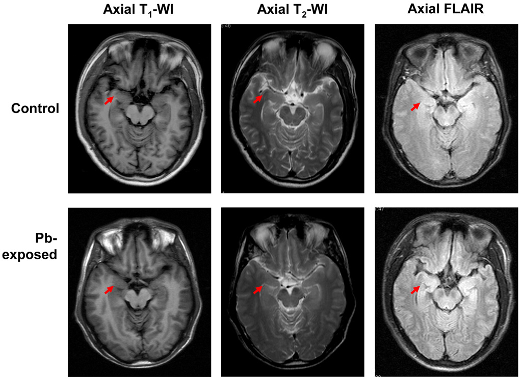
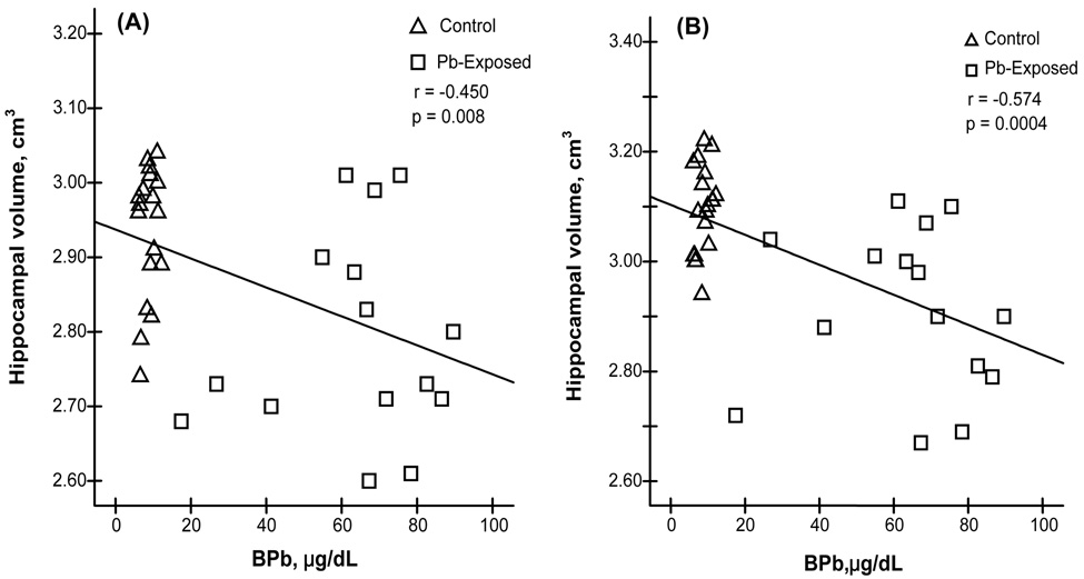
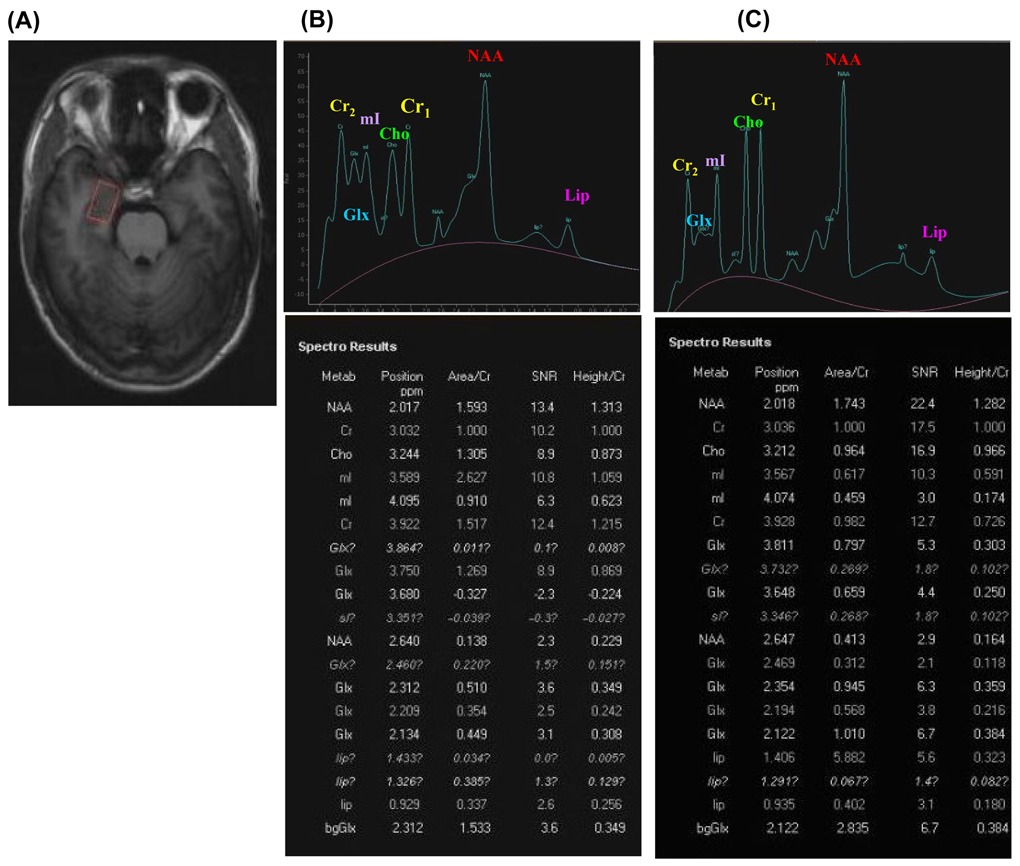
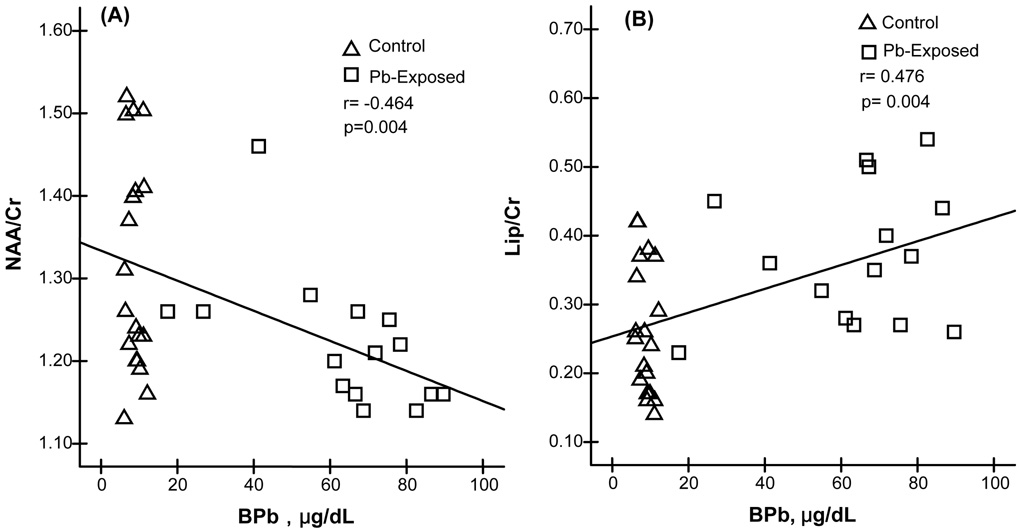
Environmental and occupational exposure to lead (Pb) remains to be a major public health issue. The purpose of this cross-sectional study was to use non-invasive magnetic resonance imaging (MRI) and proton magnetic resonance spectroscopy ((1)H MRS) techniques to investigate whether chronic exposure to Pb in an occupational setting altered brain structure and function of Pb-exposed workers. The Pb-exposed group consisted of 15 workers recruited from either a Pb-smelting factory or a Pb-battery manufacturer. The control group had 19 healthy volunteers who had no history of Pb exposure in working environment or at home. The average airborne Pb concentrations in fume and dust were 0.43 and 0.44 mg/m(3), respectively, in the smeltery, and 0.10 and 1.06 mg/m(3), respectively, in the Pb battery workshop. The average blood Pb concentrations (BPb) in Pb-exposed and control workers were 63.5 and 8.7 microg/dL, respectively. The MRI examination showed that brain hippocampal volume among Pb-exposed workers was significantly diminished in comparison to age-matched control subjects (p < 0.01), although the extent of this reduction was relatively small (5-6% of the control values). Linear regression analyses revealed significant inverse associations between BPb and the decreased hippocampal volume on both sides of brain hemisphere. Among five brain metabolites investigated by MRS, i.e., N-acetyl-aspartate (NAA), creatine (Cr), choline (Cho), inosine (mI), glutamate/glutamine (Glx) and lipids (Lip), a significant decrease in NAA/Cr ratio (7% of controls, p < 0.05) and a remarkable increase in Lip/Cr ratio (40%, p < 0.01) were observed in the brains of Pb-exposed workers as compared to controls. Furthermore, the increased Lip/Cr ratio was significantly associated with BPb (r = 0.46, p < 0.01). Taken together, this study suggests that occupational exposure to Pb may cause subtle structural and functional alteration in human brains. The MRI and MRS brain imaging techniques can be used as the non-invasive means to evaluate Pb-induced neurotoxicity.
Figures
References
-
- Alfano DP, Petit TL. Neonatal lead exposure alters the dendritic development of hippocampal dentate granule cells. Exp Neurol. 1982;75:275–288. - PubMed
-
- Antuono PG, Jones JL, Wang Y, Li SJ. Decreased glutamate + glutamine in Alzheimer’s disease detected in vivo with (1) H-MRS at 0.5T. Neurology. 2001;56:737–742. - PubMed
-
- Barbosa F, Jr, Ramires I, Rodrigues MH, Saint' Pierre TD, Curtius AJ, Buzalaf MR, Gerlach RF, Tanus-Santos JE. Contrasting effects of age on the plasma/whole blood lead ratio in men and women with a history of lead exposure. Environ Res. 2006;102:90–95. - PubMed
-
- Bennet C, Bettaiya R, Rajanna S, Baker L, Yallapragada PR, Brice JJ, White SL, Bokara KK. Region specific increase in the antioxidant enzymes and lipid peroxidation products in the brain of rats exposed to lead. Free Rad Res. 2007;41:267–273. - PubMed
-
- Bernasconi N, Bernasconi A, Caramanos Z, Antel SB, Andermann F, Arnold DL. Mesial temporal damage in temporal lobe epilepsy: A volumetric MRI study of the hippocampus, amygdala and parahippocampal region. Brain. 2003;126:462–469. - PubMed
Publication types
MeSH terms
Substances
Grants and funding
LinkOut - more resources
Full Text Sources
Medical
