. 2008 Sep 30;379(2):284-93.
doi: 10.1016/j.virol.2008.07.006. Epub 2008 Aug 8.

Acute cellular uptake of abnormal prion protein is cell type and scrapie-strain independent

Affiliations

Acute cellular uptake of abnormal prion protein is cell type and scrapie-strain independent

Christopher S Greil et al. Virology. .

Abstract

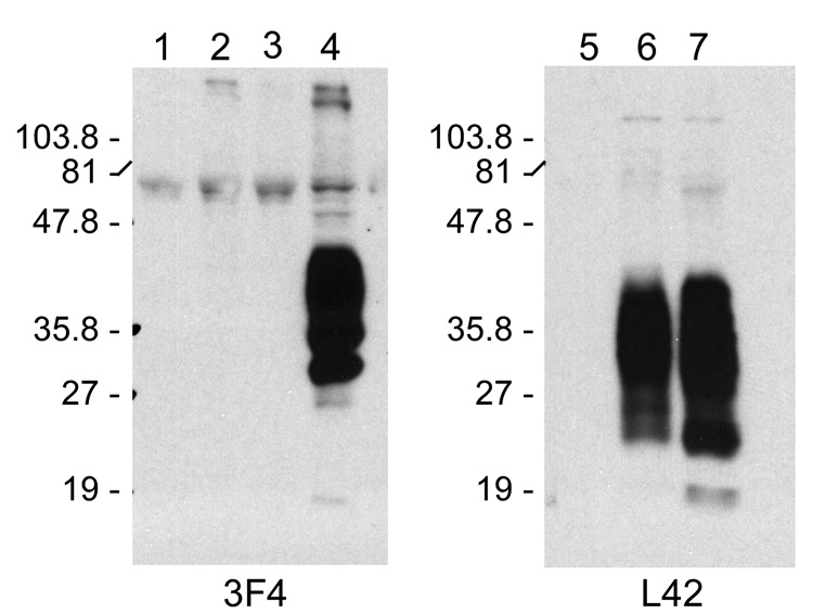
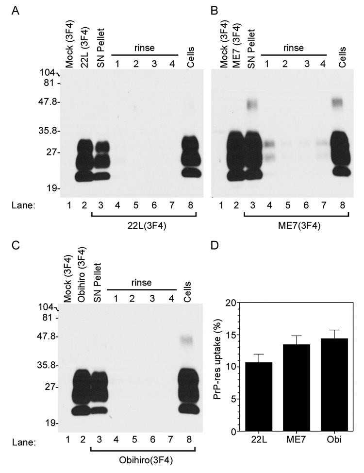
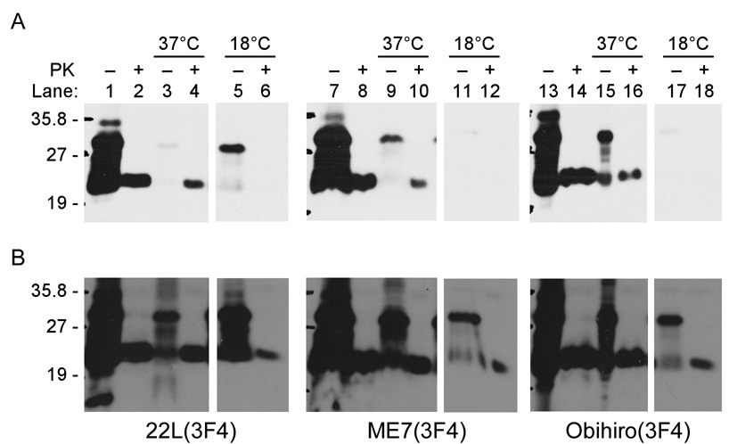
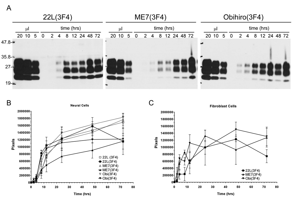
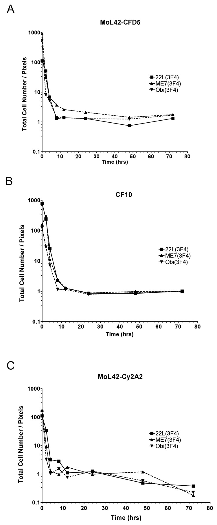
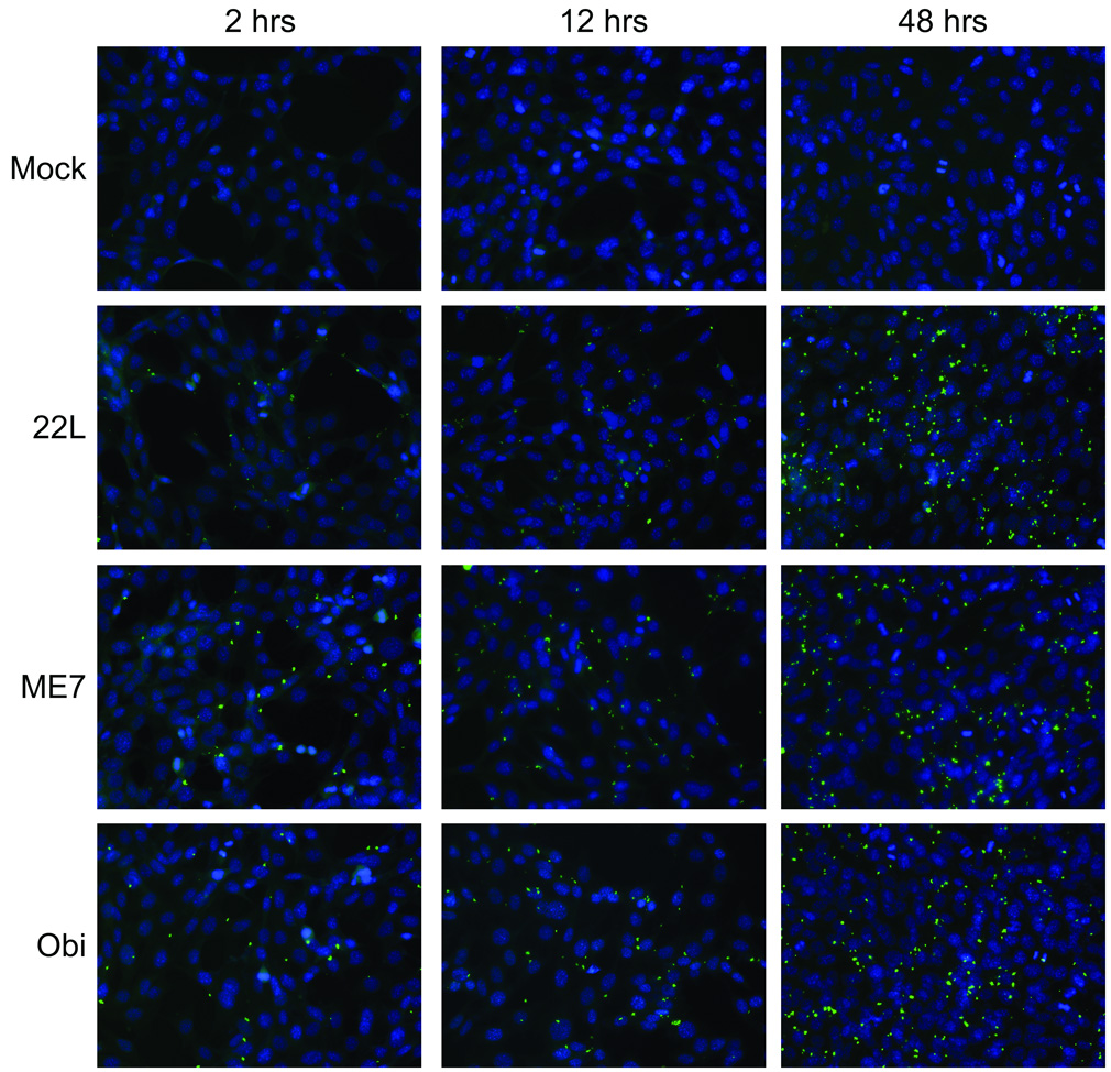
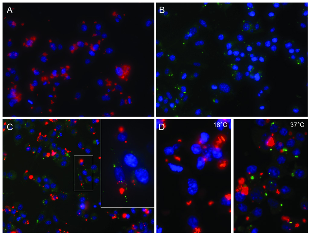
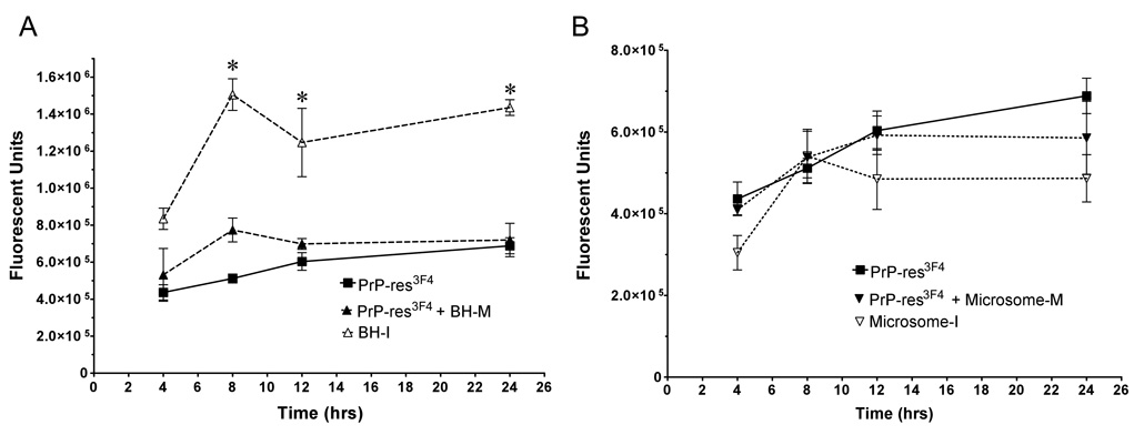
Transmissible spongiform encephalopathies (TSEs) are fatal neurodegenerative diseases that include Creutzfeldt-Jakob disease, bovine spongiform encephalopathy and sheep scrapie. Although one of the earliest events during TSE infection is the cellular uptake of protease resistant prion protein (PrP-res), this process is poorly understood due to the difficulty of clearly distinguishing input PrP-res from either PrP-res or protease-sensitive PrP (PrP-sen) made by the cell. Using PrP-res tagged with a unique antibody epitope, we examined PrP-res uptake in neuronal and fibroblast cells exposed to three different mouse scrapie strains. PrP-res uptake was rapid and independent of scrapie strain, cell type, or cellular PrP expression, but occurred in only a subset of cells and was influenced by PrP-res preparation and aggregate size. Our results suggest that PrP-res aggregate size, the PrP-res microenvironment, and/or host cell-specific factors can all influence whether or not a cell takes up PrP-res following exposure to TSE infectivity.


Figures

Figure 1
Figure 1. Cell line expression of PrP
Western blot detection of PrP-sen with either mouse monoclonal antibody 3F4 (left panel) or mouse monoclonal antibody L42 (right panel) for cell lines CF10 (lanes 1, 5), MoL42-CFD5 (lanes 2, 6), and MoL42-Ψ2A2 (lanes 3, 7). As a positive control for the specificity of the 3F4 antibody, Lane 4 shows CF10 cells expressing Mo3F4.
Figure 2
Figure 2. Mouse neural cells take up PrP-res3F4
Western blot analysis of PrP-res3F4 uptake into MoL42-CFD5 cells. Brain homogenate from 22L, ME7 or Obihiro scrapie-infected Tg(WT-E1) mice were PK treated and used as positive controls (Panels A–C, lane 2) while PK treated brain homogenate from mock-infected Tg(WT-E1) mice was used as negative controls (Panel A–C, lane 1). For both the positive and negative controls, 1/20th of the total homogenate used was loaded onto the gel. Brain homogenates (200µl of a 1% brain homogenate) from 22L(3F4) (panel A), ME7(3F4) (panel B), or Obi(3F4) (panel C) were added to MoL42-CFD5 cells for 24 hours and then removed from the cell monolayer. Cell debris and dead cells were spun out of the supernatant and the supernatant pellet was PK treated, methanol precipitated and the entire sample loaded onto the gel (SN Pellet). Cells were then rinsed with PBS four times (rinse 1–4), lysed, and both the cell lysate and rinses were PK treated, methanol precipitated and the entire sample loaded onto the gel. PrP-res3F4 was detected in the lysed and PK treated cells (Cells). All blots were analyzed using the mouse monoclonal antibody 3F4 and developed using ECL (Amersham). The percentage of total PrP-res3F4 taken up by the cells after 24 hours is shown in Panel D. PrP-res3F4 was quantified as detailed in the materials and methods. There was no significant difference between the three strains in the amount of PrP-res taken up by the MoL42-CFD5 cells (p>0.05 using 1-way Anova with Bonferroni’s multiple comparison test). Data are shown as mean ± S.D. for N=12.
Figure 3
Figure 3. PrP-res3F4 uptake into neural cells is significantly decreased at 18°C
MoL42-CFD5 cells were exposed to infected brain homogenate from 22L(3F4), ME7(3F4) or Obi(3F4) scrapie for 24 hours at either 37°C or 18°C. Samples were PNGaseF treated to remove complex glycans. Non-PK digested infected brain homogenates were run to illustrate total PrP levels (lanes 1,7,13) while PK-digested brain homogenates were run as a positive control for PrP-res (lanes 2,8,14). Some PrP-sen3F4 was taken up by cells at 37°C (−PK lanes 3, 9, 15) and PrP-res3F4 was detected in cells which had been exposed to scrapie brain homogenate and incubated at 37°C (+PK lanes 4, 10, 16). Exposure time for Panel A was 8 minutes. After overnight exposures of the gels, some PrP-sen3F4 (−PK lanes 5, 11, 17) and a low level of PrP-res3F4 (+PK lanes 6, 12, 18) were detected in cells which had been exposed to scrapie brain homogenate and incubated at 18°C (Panel B). All blots were analyzed using the mouse monoclonal antibody 3F4 and developed using ECL (Amersham).
Figure 4
Figure 4. Kinetics of PrP-res3F4 uptake into cells
A) Representative Western blot analysis of PrP-res3F4 uptake into MoL42-CFD5 cells from 0 – 72 hrs. Scrapie-infected brain homogenates from 22L(3F4), ME7(3F4) or Obi(3F4) were analyzed. The first three lanes of each gel show PK-treated scrapie-infected brain homogenate for the appropriate strain loaded in 20 ul, 10 ul, and 5 ul volumes. These lanes were used as internal standards to establish a constant film exposure time from experiment to experiment (4min) in order to quantify the data within the linear range of the film. The amount of PrP-res internalized by the cells increased over time. B) Graphical representation of total cell-associated 22L(3F4), ME7(3F4), or Obi(3F4) taken up by either MoL42-CFD5 cells (solid lines N=5 or 6) or CF10 cells (dotted lines N=3) from 0–72 hrs. The error bars represent SEM. No statistically significant differences were found between the scrapie strains (p>0.05, Two-way ANOVA) or between the different cell types (p>0.05, Mann Whitney test) for each scrapie strain. C) Graphical representation of total cell-associated 22L, ME7 or Obihiro PrP-res3F4 taken up by MoL42-ψ2A2 fibroblast cells (N=3) from 0 – 72 hrs where the error bars represent SEM. No statistically significant differences were found between the scrapie strains (p>0.05, Two-way ANOVA) or between the fibroblast and neuronal cell types (p>0.05, Mann Whitney test). All blots were analyzed using the mouse monoclonal antibody 3F4. For all data, the amount of PrP-res is expressed in pixels and was quantified by analysis of ECL developed western blots using the UN-SCAN-IT software as detailed in the Methods.
Figure 5
Figure 5. Increased levels of cellular PrP-res3F4 correlate with increased cell number
Ratio of total cell number to total cell-associated PrP-res3F4 for scrapie strains 22L(3F4), ME7(3F4) or Obi(3F4). PrP-res3F4 was quantified using ECL developed western blots analyzed with the UN-SCAN-IT software. A) Ratio of the number of MoL42-CFD5 cells to total PrP-res3F4 (N=5). B) Ratio of the number of CF10 cells to total PrP-res3F4 (N=6). C) Ratio of the number of MoL42-ψ2A2 cells to total PrP-res3F4 (N=6). The variability of the data at later time points was likely due to increased cell death over time (data not shown) rather than any cell-type differences.
Figure 6
Figure 6. PrP-res3F4 uptake into neural cells using immunofluorescence microscopy
PrP-res3F4 partially purified from 22L(3F4), ME7(3F4), Obi(3F4) or mock brain homogenates was added to MoL42-CFD5 cells for 2 – 48 hrs. Cells were rinsed, fixed and immunolabeled with the mouse monoclonal antibody 3F4. Anti-mouse FITC labeled antibody (green) was used to detect PrP-res3F4 while DAPI stain (blue) denotes cell nuclei. All images were taken with a 40X objective.
Figure 7
Figure 7. Large PrP-res aggregates bind to the cell surface and are not rapidly internalized
Differentiated SN56 cells were exposed to Alexa-Fluor-596 labeled (red) or unlabeled (green) 22L PrP-res3F4 for 24 hours. Cells were then rinsed, fixed and when necessary, immunolabeled with the mouse monoclonal antibody 3F4 (Panels B–D). Anti-mouse FITC labeled antibody (green) was used to detect 22L PrP-res3F4, while DAPI stain (blue) denotes cell nuclei. Alexa-Fluor-596 labeled proteins are red. All images were taken with a 40X objective. Cells were exposed to A) Alexa-Fluor labeled partially purified 22L PrP-res3F4 (red), B) partially purified 22L PrP-res3F4 (green), C) Alexa- Fluor labeled partially purified PrP-res from a 22L scrapie-infected wild type mouse (red) and partially purified 22L PrP-res3F4 (green) (inset taken with a 60X objective). In panel D, cells were exposed for 24 hours at either 18°C or 37°C to Alexa-Fluor labeled partially purified PrP-res from a 22L scrapie-infected wild type mouse (red) and 22L PrP-res3F4 (green). The larger Alexa-Fluor labeled PrP-res aggregates do not colocalize with the smaller, more punctate PrP-res3F4 aggregates.
Figure 8
Figure 8. Infectious brain homogenate PrP-res3F4 is taken up more efficiently then either microsome or partially purified PrP-res3F4
Graphical representation of PrP-res3F4 uptake from 0 – 24 hrs into MoL42-CFD5 cells (N = 6) where error bars represent SEM. A) Kinetics of PrP-res3F4 uptake using either infectious brain homogenate PrP-res3F4 (BH-I), partially purified PrP-res3F4 (PrP-res3F4) or partially purified PrP-res3F4 with mock infected brain homogenate (PrP-res3F4 + BH-M) added to match the total protein content of BH-I (* p<0.05, t = 8–24 hrs, Bonferroni test). B) Kinetics of PrP-res3F4 uptake using either infectious microsome PrP-res3F4 (Microsome-I), purified PrP-res3F4 (PrP-res3F4) or partially purified PrP-res3F4 with mock infected microsomes (PrP-res3F4 + Microsome-M) added to match total protein content of infectious microsomes (p>0.05 for all time points, Bonferroni test). All data was obtained with IR-dye 800CW developed western blots where the PrP-res3F4 level was quantified as fluorescent units using the Li-Cor Odyssey imaging system and associated software.

References

    1. Baron GS, Magalhaes AC, Prado MA, Caughey B. Mouse-adapted scrapie infection of SN56 cells: greater efficiency with microsome-associated versus purified PrP-res. J. Virol. 2006;80:2106–2117. - PMC - PubMed
    1. Baron GS, Wehrly K, Dorward DW, Chesebro B, Caughey B. Conversion of raft associated prion protein to the protease-resistant state requires insertion of PrP-res (PrP(Sc)) into contiguous membranes. EMBO J. 2002;21:1031–1040. - PMC - PubMed
    1. Bendheim PE, Barry RA, DeArmond SJ, Stites DP, Prusiner SB. Antibodies to a scrapie prion protein. Nature. 1984;310:418–421. - PubMed
    1. Beranger F, Mange A, Goud B, Lehmann S. Stimulation of PrP(C) retrograde transport toward the endoplasmic reticulum increases accumulation of PrP(Sc) in prion-infected cells. J.Biol.Chem. 2002;277:38972–38977. - PubMed
    1. Bergstrom AL, Jensen TK, Heegaard PM, Cordes H, Hansen VB, Laursen H, Lind P. Short-term study of the uptake of PrP(Sc) by the Peyer's patches in hamsters after oral exposure to scrapie. J.Comp Pathol. 2006;134:126–133. - PubMed

Publication types