

Review
doi: 10.1016/j.cell.2008.07.025.
TGF-beta: a master of all T cell trades
Affiliations
- PMID: 18692464
- PMCID: PMC3677783
- DOI: 10.1016/j.cell.2008.07.025
Item in Clipboard
Review
TGF-beta: a master of all T cell trades
Cell.
.
Abstract
A functional adaptive immune system depends on a diverse and self-tolerant population of T lymphocytes that are generated in the thymus and maintained in the peripheral lymphoid organs. Recent studies have defined the cytokine transforming growth factor-beta (TGF-beta) as a critical regulator of thymic T cell development as well as a crucial player in peripheral T cell homeostasis, tolerance to self antigens, and T cell differentiation during the immune response. The unique mechanism of TGF-beta activation and the plasticity of TGF-beta signaling create a stage for TGF-beta to integrate signals from multiple cell types and environmental cues to regulate T cells.
Figures
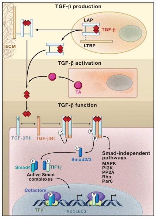
TGF-β is synthesized in an inactive form composed of a TGF-β dimer in association with the latency-associated protein (LAP). This latent TGF-β molecule can be secreted as such, or can form a complex with latent-TGF-β-binding protein (LTBP) that mediates its deposition to the extracellular matrix (ECM). TGF-β becomes activated after the engagement of a TGF-β activator (TA) that triggers LAP degradation or alters LAP’s conformation in response to environmental cues. Active TGF-β binds to a tetrameric complex composed of TGF-β receptor II (TGF-βRII) and TGF-β receptor I (TGF-βRI) and initiates signaling pathways that are dependent on the kinase activity of the receptors. Activated TGF-βRI phosphorylates the transcription factors Smad2 and Smad3, triggering their translocation into the nucleus in complex with the proteins Smad4 or TIF1γ. Smad complexes in association with additional transcription factors (TFs) bind to the regulatory sequences in target genes and regulate gene expression by recruiting transcription cofactors. In addition, TGF-β activates Smad-independent pathways such as those mediated by mitogen-activated protein kinase (MAPK), PI3K kinase, PP2A phosphatase, Rho family proteins, and the epithelial polarity protein Par6, which trigger different cell type-specific responses.
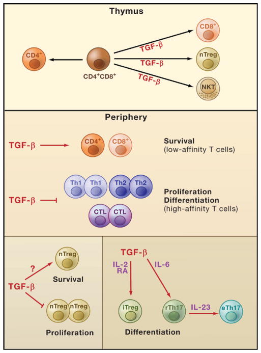
The cytokine TGF-β regulates T cell development, homeostasis, tolerance, and immunity. TGF-β signaling in T cells promotes differentiation of thymic T cells into natural killer T (NKT) cells, natural regulatory T (nTreg) cells, and CD8+ T cells. In peripheral tissues, TGF-β signaling in T cells is likely to be essential for the survival of low-affinity CD4+ and CD8+ T cells. It also modulates immune tolerance by inhibiting high-affinity CD4+ and CD8+ T cell proliferation and differentiation into T helper 1 (Th1), Th2, and cytotoxic T lymphocytes (CTL). In addition, TGF-β signaling in nTreg cells inhibits their proliferation but supports their maintenance in peripheral lymphoid organs, a process likely mediated by TGF-β control of nTreg cell survival. TGF-β also positively regulates differentiation of certain peripheral T cells in conjunction with other factors. TGF-β promotes the differentiation of induced Treg (iTreg) cells that is potentiated by the cytokine interleukin-2 (IL-2) and retinoic acid (RA). In the presence of IL-6, TGF-β drives the differentiation of Th17 cells and maintains them in a regulatory state (rTh17). Stimulation of rTh17 cells by IL-23 in the absence of TGF-β enables these cells to acquire effector functions and promotes their differentiation into effector Th17 cells (eTh17).
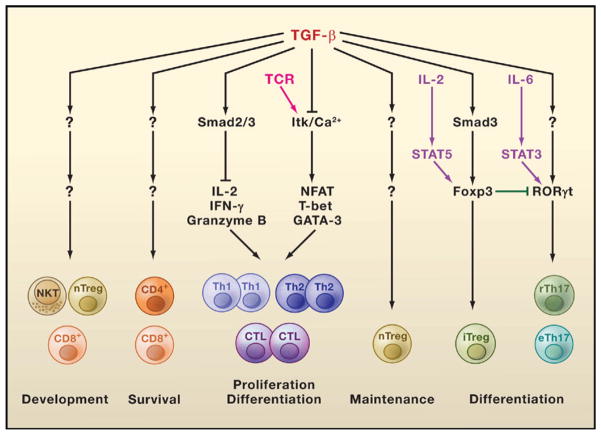
The Smad2/3 complexes are involved in TGF-β inhibition of interleukin-2 (IL-2), interferon-γ (IFN-γ), and granzyme B transcription required for effector T helper 1 (Th1), Th2, and cytotoxic T lymphocyte (CTL) proliferation and differentiation. In addition, TGF-β blocks T cell receptor (TCR)-induced Tec kinase Itk activation and Ca2+ responses that are required for the expression of other transcription factors such as NFAT, T-bet, and GATA-3, which are also involved in T cell activation and differentiation. Induced regulatory T (iTreg) cell differentiation triggered by TGF-β occurs via Smad3-dependent induction of Foxp3 expression, which is enhanced by activation of the transcription factor STAT5 (induced by the cytokine IL-2). TGF-β induction of Th17 cell differentiation is associated with its induction of RORγt expression, which is potentiated by the IL-6-activated transcription factor STAT3. The transcription factor Foxp3 interacts with the transcription factor RORγt and suppresses its function, providing a mechanism for the reciprocal differentiation of iTreg and Th17 cells. The mechanisms of TGF-β-induced development of natural regulatory T (nTreg) cells, natural killer T (NKT) cells, and CD8+ T cells are unknown. How TGF-β regulates the survival of low-affinity CD4+ and CD8+ T cells and maintains nTreg cells is also unknown.
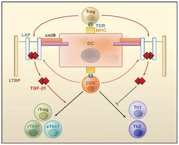
Upon recognition of antigens presented by dendritic cells (DCs), Treg cells are activated and secrete the latent form of TGF-β1. This latent form comprises a TGF-β1 dimer associated with the latency-associated protein (LAP), which may additionally recruit latent TGF-β-binding protein (LTBP). Interaction of LAP with αvβ8 integrin expressed by DCs triggers degradation of LAP (mediated by un-identified proteases, not shown) and the release of the active form of TGF-β1. Active TGF-β1 subsequently acts on naive CD4+ T cells via a paracrine mechanism to inhibit their differentiation into Th1 or Th2 cells and to promote their differentiation into induced regulatory T (iTreg) cells, regulatory Th17 (rTh17) cells, or effector Th17 (eTh17) cells. Activated CD4+ T cells can also produce low amounts of latent TGF-β1 that potentially regulate T cell differentiation through an autocrine route.
References
-
- Annes JP, Munger JS, Rifkin DB. Making sense of latent TGFbeta activation. J Cell Sci. 2003;116:217–224. - PubMed
-
- Bendelac A, Rivera MN, Park SH, Roark JH. Mouse CD1-specific NK1 T cells: Development, specificity, and function. Annu Rev Immunol. 1997;15:535–562. - PubMed
-
- Bettelli E, Carrier Y, Gao W, Korn T, Strom TB, Oukka M, Weiner HL, Kuchroo VK. Reciprocal developmental pathways for the generation of pathogenic effector TH17 and regulatory T cells. Nature. 2006;441:235–238. - PubMed
-
- Blobe GC, Schiemann WP, Lodish HF. Role of transforming growth factor beta in human disease. N Engl J Med. 2000;342:1350–1358. - PubMed
Publication types
MeSH terms
Substances
Grants and funding
LinkOut - more resources
Full Text Sources
Other Literature Sources
