. 2008 Aug 8;134(3):427-38.
doi: 10.1016/j.cell.2008.06.022.

A network of nuclear envelope membrane proteins linking centromeres to microtubules

Affiliations

A network of nuclear envelope membrane proteins linking centromeres to microtubules

Megan C King et al. Cell. .

Abstract

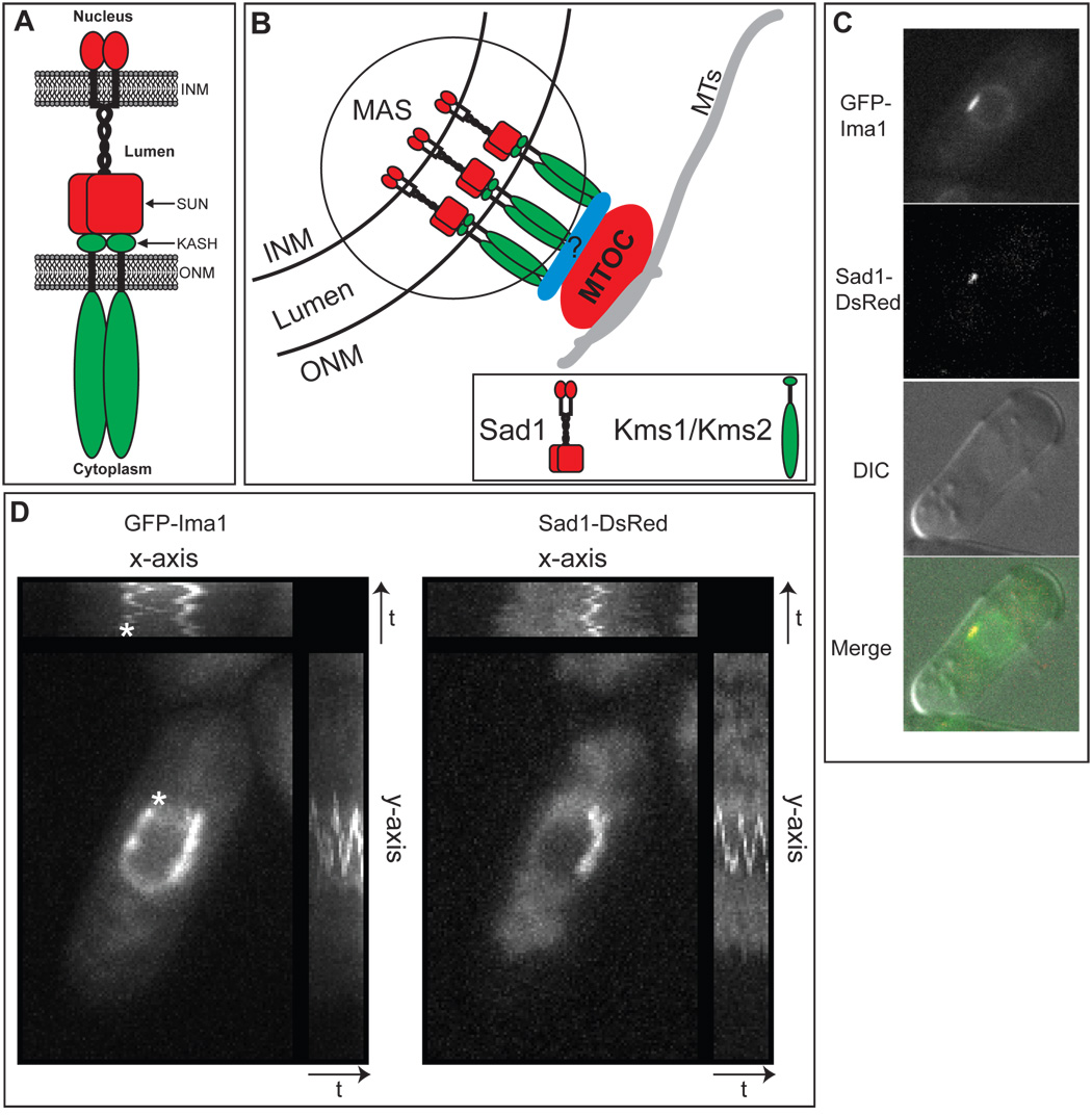
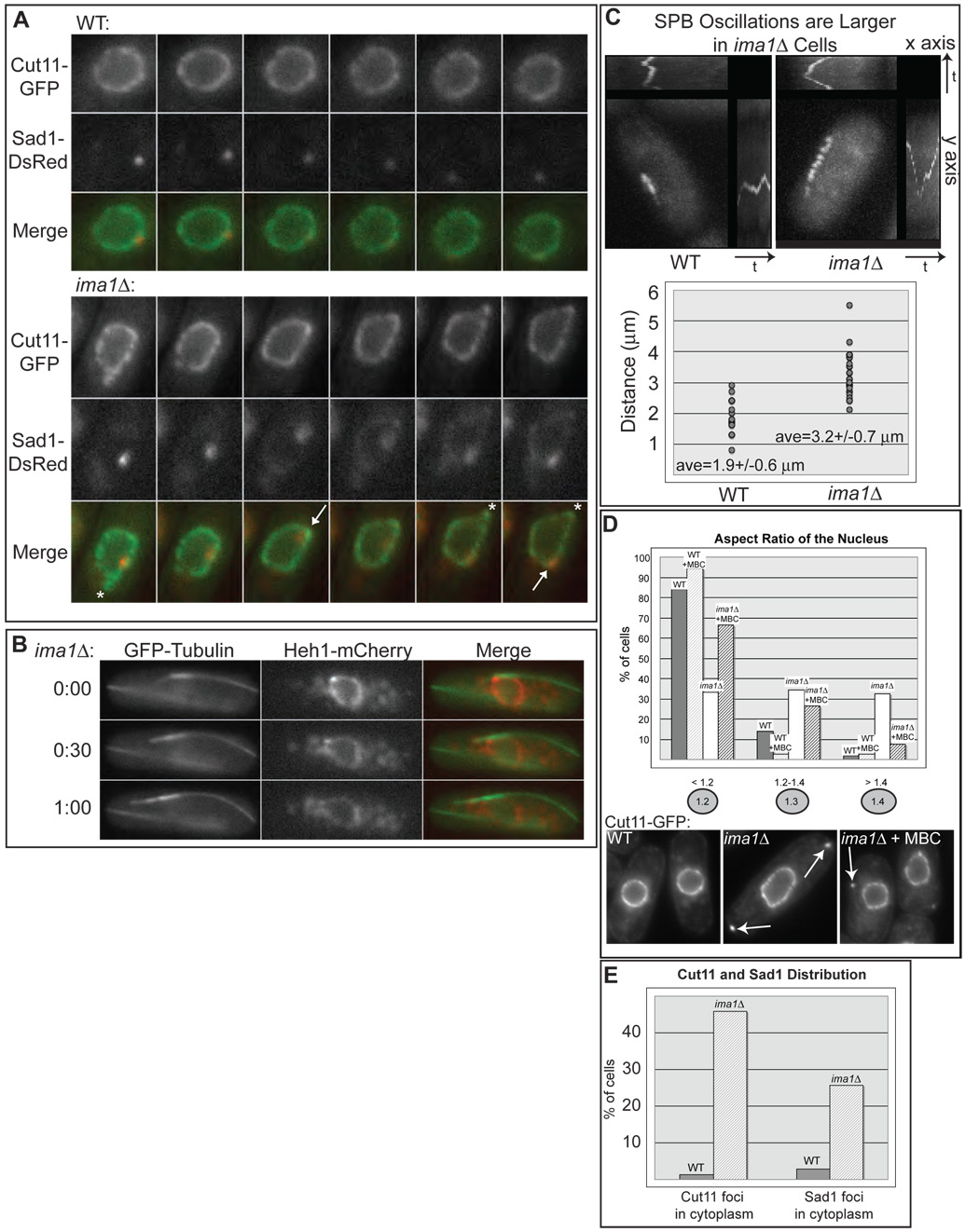
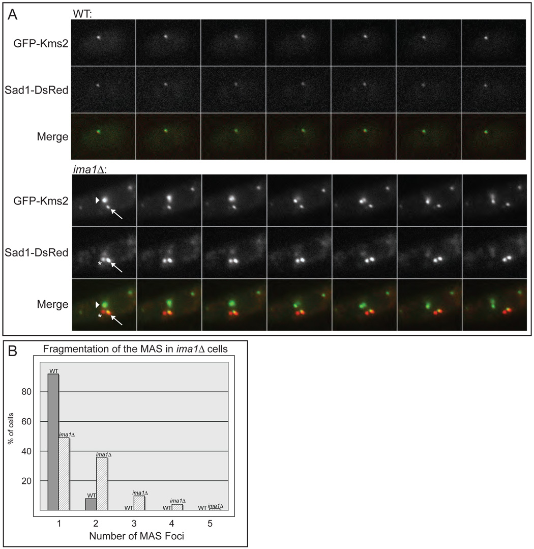
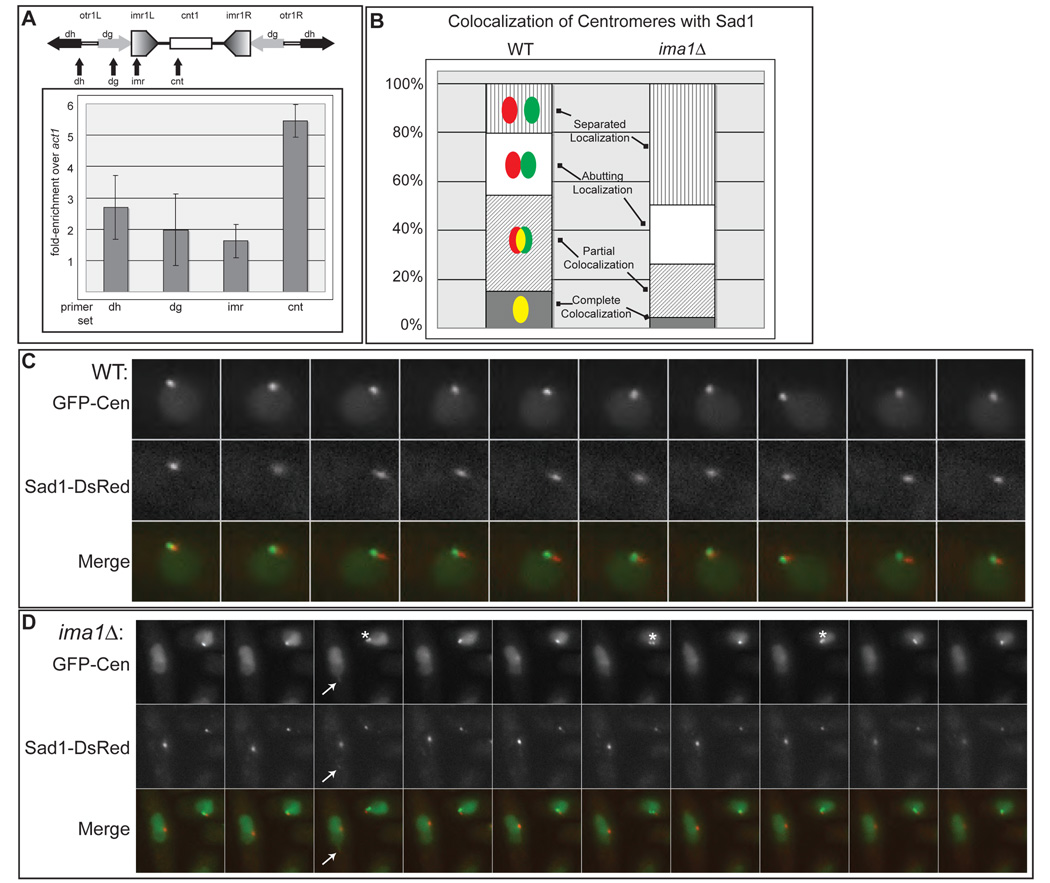
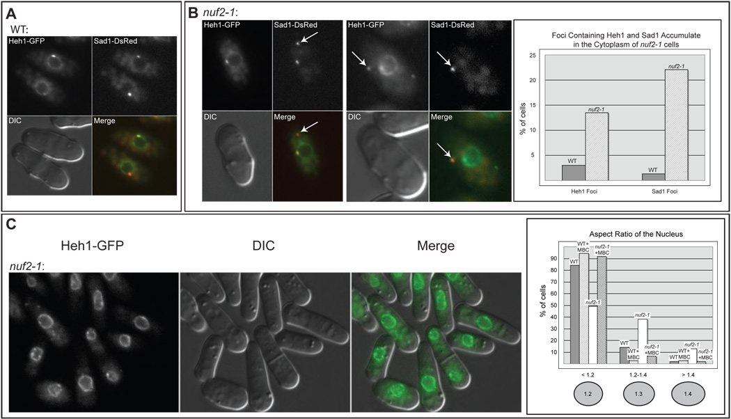
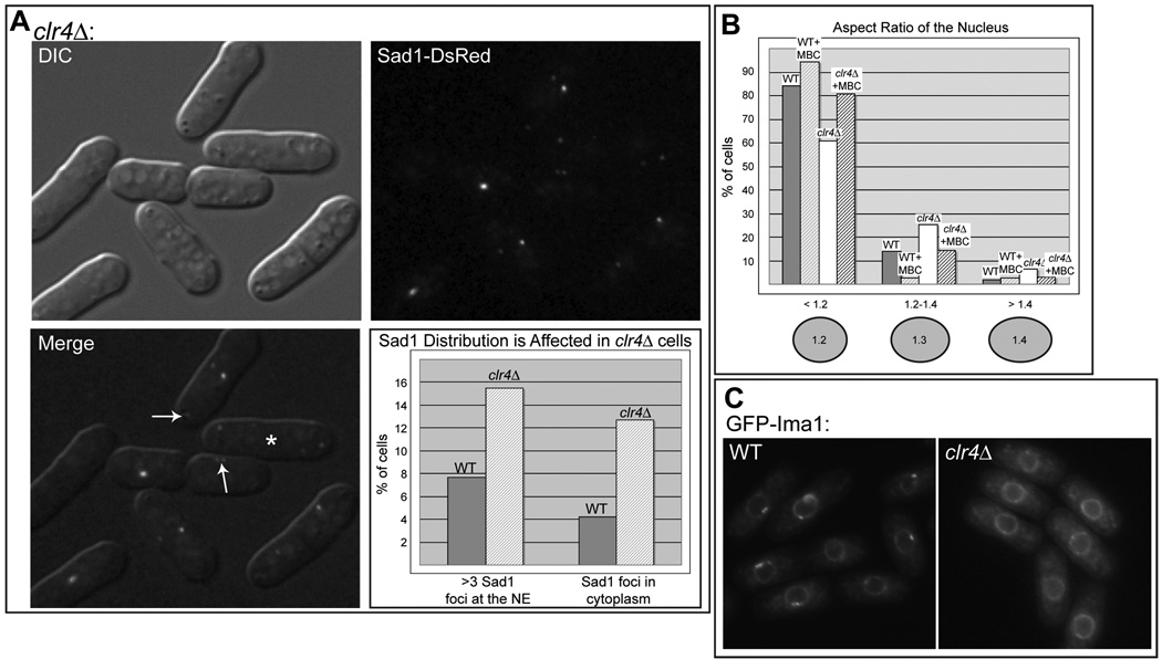
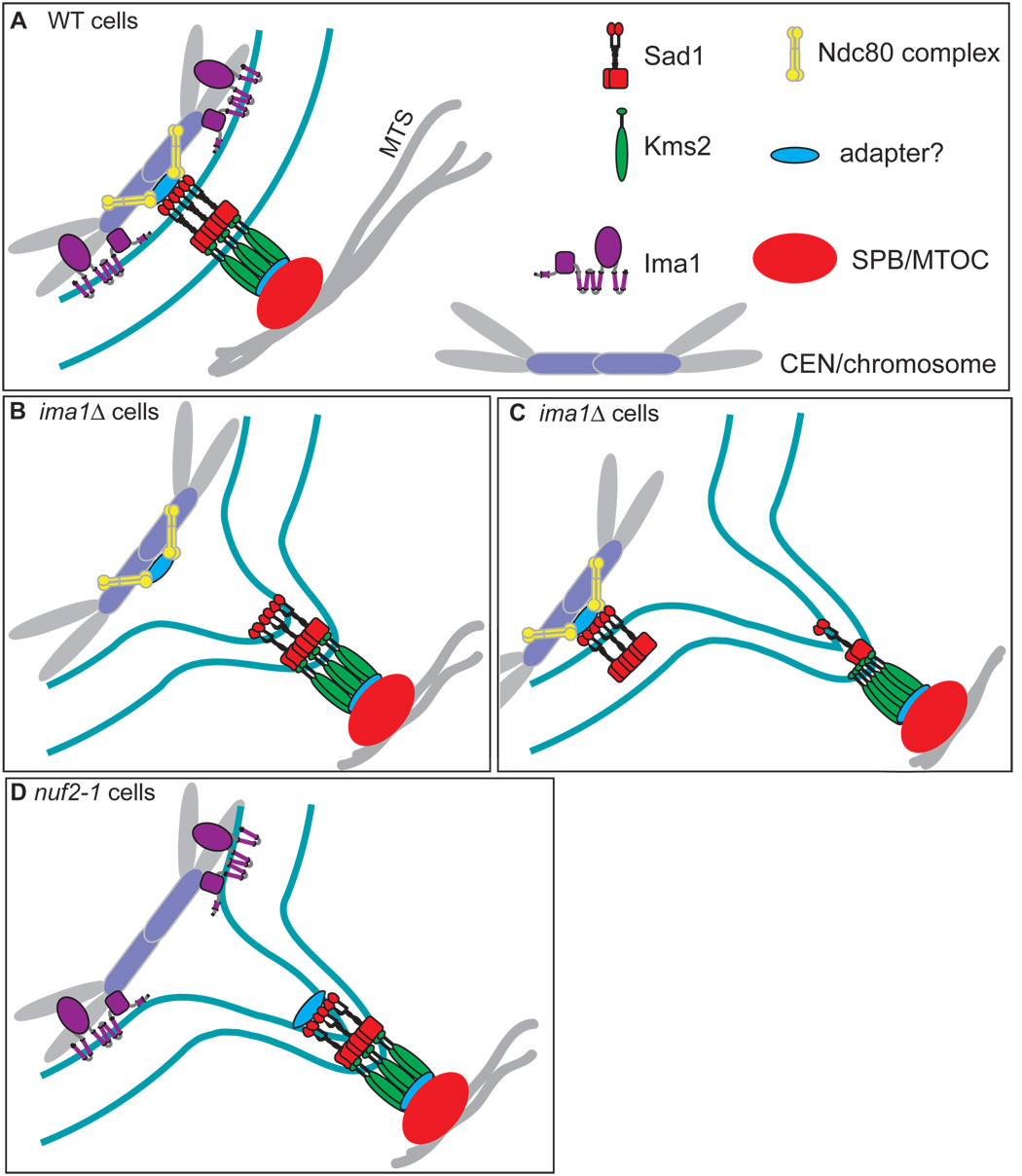
In the fission yeast S. pombe, nuclei are actively positioned at the cell center by microtubules. Here, we show that cytoplasmic microtubules are mechanically coupled to the nuclear heterochromatin through proteins embedded in the nuclear envelope. This includes an integral outer nuclear membrane protein of the KASH family (Kms2) and two integral inner nuclear membrane proteins, the SUN-domain protein Sad1 and the previously uncharacterized protein Ima1. Ima1 specifically binds to heterochromatic regions and promotes the tethering of centromeric DNA to the SUN-KASH complex. In the absence of Ima1, or in cells harboring mutations in the centromeric Ndc80 complex, inefficient coupling of centromeric heterochromatin to Sad1 leads to striking defects in the ability of the nucleus to tolerate microtubule-dependent forces, leading to changes in nuclear shape, loss of spindle pole body components from the nuclear envelope, and partial dissociation of SUN-KASH complexes. This work highlights a framework for communication between cytoplasmic microtubules and chromatin.


Figures

Figure 1
Figure 1. Ima1 is a conserved, integral inner nuclear membrane protein that is enriched at the site of MTOC attachment
A. Cartoon of SUN-KASH interactions at the nuclear envelope (NE). The SUN domain protein (red) is integrated into the inner nuclear membrane (INM). The protein has an N-terminal nucleoplasmic domain followed by a single transmembrane segment, a lumenal coiled-coil region, and the conserved SUN domain. The KASH domain protein (green) is integrated into the outer nuclear membrane (ONM). The variable N-terminal cytoplasmic domain interacts with cytoskeletal elements and the C-terminus contains the KASH domain, which is composed of the transmembrane segment (black) and a small lumenal tail (labeled KASH). Both proteins are shown as homodimers. B. Diagram of the microtubule organizing center (MTOC) attachment site (MAS) at the NE of S. pombe (circled). SUN domain protein Sad1 (red) and KASH domain proteins Kms1 and/or Kms2 (green) interact within the lumen of the NE to link the MTOC to the NE either directly or through an as yet unidentified adapter protein(s) (blue). MTs = microtubules. C. GFP-Ima1 localizes to the NE and MAS. Fluorescent micrographs, DIC and merged images are shown of a representative single cell of strain MKSP58 expressing GFP-Ima1 and Sad1-DsRed. D. GFP-Ima1 comigrates with Sad1-DsRed as the SPB oscillates along the NE. A composite image of time-lapse frames taken every 15 seconds for 5 minutes of one MKSP58 cell (see above) expressing GFP-Ima1and Sad1-DsRed. The asterisk indicates a second focus of GFP-Ima1 that oscillates along the NE. The time axes are indicated by the arrow and “t”.
Figure 2
Figure 2. ima1Δ cells have NE and MAS defects
A. Large deformations of the NE in the absence of Ima1. Fluorescent micrographs of one WT (top panels) and one ima1Δ cell (bottom panels) expressing Cut11-GFP and Sad1-DsRed (MKSP50 and MKSP56, respectively) taken in consecutive 60 second intervals are shown along with the merged images. Deformations led by Sad1-DsRed are labeled with arrows; deformations without visible Sad1-DsRed are labeled with asterisks. B. Large deformations of the NE in ima1Δ cells are driven by MTs. Fluorescent micrographs of one ima1Δ cell expressing GFP-tubulin and Heh1-mCherry (MKSP173) imaged every 30 seconds for one minute (time indicated on the left) and shown with the merged images. C. SPB oscillations are larger in ima1Δ cells. Composite images of time-lapse frames taken every 15 seconds for 5 minutes of one WT cell (MKSP81) and one ima1Δ cell (MKSP152) expressing GFPKms2 (upper panel). Plot of the maximum SPB oscillation amplitude for size-matched WT and ima1Δ cells (n=25, lower panel). The average value (ave) is given with its standard deviation. D. MT forces lead to defects in nuclear shape and cytoplasmic Cut11-GFP foci appear (arrows) in ima1Δ cells. Quantitation of the spherical nature of WT (MKSP10) versus ima1Δ (MKSP22) nuclei. As indicated, WT and ima1Δ cells were also treated for 15 minutes with carbendazim (MBC) prior to imaging. Measurements are presented from at least 100 non-mitotic cells. E. Percentage of WT (MKSP50) and ima1Δ (MKSP56) cells that display Cut11-GFP and Sad1-DsRed cytoplasmic foci. n = >100 interphase cells.
Figure 3
Figure 3. Sad1 and Kms2 become disorganized in the absence of Ima1
A. Top panel. Fluorescence micrographs demonstrate that the KASH domain protein GFP-Kms2 and Sad1-DsRed colocalize at the MAS in WT cells (MKSP81). Time-lapse images taken of one cell every 30 seconds for 3 minutes are shown. Bottom panel. GFP-Kms2 and Sad1-DsRed fragment into multiple foci at the NE and no longer colocalize stoichiometrically in ima1Δ cells. Fluorescence micrographs of one cell of strain MKSP152 taken every 30 seconds for 3 minutes. Arrow indicates a focus with both GFP-Kms2 and Sad1-DsRed; arrowhead indicates a focus with only GFP-Kms2; asterisk indicates a focus with only Sad1-DsRed (see text). B. Quantitation of the number of MAS foci at the NE in WT and ima1Δ cells. Strains (MKSP81 and MKSP152) were analyzed for the total number of foci containing GFP-Kms2 and/or Sad1-DsRed in non-mitotic cells. n = >100 cells.
Figure 4
Figure 4. Centromeres becomes uncoupled from the MAS in ima1Δ cells
A. GFPIma1 associates with centromeres. Schematic representation of the Chromosome I centromere. otr=outer repeat, imr=innermost repeat, cnt1=center region, dg=dg repeats, dh=dh repeats. Arrows indicate positions of primer sets used in real-time PCR analysis. Graph of fold-enrichment of GFP-Ima1 by chromatin immunoprecipitation at the given region over the control actin gene location, act1. The average of three independent immunoprecipitations are presented with their standard deviations. B. Centromeres are separated from Sad1-DsRed in ima1Δ cells. For each cell, the degree of colocalization of LacI-GFP associated with Lac operators integrated into centromere II (cen2:lacOp, green, labeled GFP-Cen) with Sad1-DsRed (red) was categorized as illustrated in the cartoons with yellow representing colocalization. The percentage of cells displaying each localization pattern was plotted. The graphs represent the average of three separate experiments consisting of greater than 100 cells each. C. In WT cells, GFP-Cen remains closely associated with the MAS during SPB oscillation. Time-lapse images of GFP-LacI and Sad1-DsRed in WT cells with integrated cen2:lacOp (MKSP71) taken every 30 seconds for 5 minutes. D. In ima1Δ cells the centromeres of sister chromatids often separate (asterisk) and no longer colocalize with Sad1-DsRed. Time-lapse images of GFP-LacI and Sad1-DsRed in an ima1Δ cell with integrated cen2:lacOp (MKSP158) were taken every 30 seconds for 5 minutes. The arrow indicates the distended nucleus (green channel) led by a weak focus of Sad1-DsRed (red channel), which is not associated with the centromeres.
Figure 5
Figure 5. Release of centromeric heterochromatin from the MAS causes NE and MAS defects
A. Heh1-GFP is an INM protein that is enriched at the MAS. Micrographs of MKSP47 cells expressing Heh1-GFP and Sad1-DsRed shown with the DIC and merged images. B. A temperature-sensitive mutation in Nuf2 leads to NE and MAS defects. Fluorescence micrographs of cells expressing Heh1-GFP and Sad1-DsRed in the nuf2-1 strain (MKSP109) at 25°C. Sad1-DsRed foci and Heh1-GFP foci are seen in the cytoplasm of nuf2-1 cells (arrows). Quantitation of Heh1 and Sad1 foci in WT versus nuf2-1 cells was determined as in Figure 2. C. The nuf2-1 mutation leads to defects in NE shape. Fluorescence micrographs of strain MKSP109 at 25°C. Quantitation of nuclear shape was carried out as in Figure 2.
Figure 6
Figure 6. Disruption of histone H3-K9 methylation leads to NE and MAS defects and mislocalization of GFP-Ima1
A. Sad1 localizes to multiple foci at the NE and to cytoplasmic foci in clr4Δ cells (MKSP123). Fluorescence micrograph showing Sad1-DsRed in the cytoplasm (top arrow). Sad1 is also found fragmented at the NE (lower arrow), including in cells that have recently completed mitosis, which have two Sad1-DsRed foci per nucleus (asterisk). Quantitation of cytoplasmic Sad1 foci was carried out as in Figure 2. B. clr4Δ cells have a mild defect in nuclear shape. Quantitation of nuclear shape was carried out as in Figure 5, using Heh1-GFP as the NE marker (MKSP119). C. The accumulation of Ima1 to the MAS is reduced in clr4Δ cells. Fluorescence micrographs of GFP-Ima1 in WT (MKSP14) and clr4Δ (MKSP120) cells.
Figure 7
Figure 7. Model of the MAS in WT, ima1Δ and nuf2-1 cells
A. In WT cells, the Sad1 (red) / Kms2 (green) bridging complex links the MTOC (SPB, orange) to the centromeric heterochromatin (lavender/gray). Ima1 (purple) resides at the inner nuclear membrane and contributes to the coupling of heterochromatin to the inner MAS. The Ndc80 complex (yellow) also participates in linking the centromeres to the inner MAS, through an unknown adapter (blue) and Sad1. B. Without Ima1, the Ndc80 complex (yellow) is not sufficient to tightly couple centromeres to the MAS, leading to large nuclear envelope (NE) deformations. C. In the absence of Ima1, fragmentation of the MAS occurs and Sad1-Kms2 lumenal interactions are disrupted, decoupling forces delivered on the NE by the SPB from the centromeres and other MAS components. D. In nuf2-1 cells, Ima1 is not sufficient to couple centromeres to the MAS, resulting in large NE deformations.

References

    1. Akhtar A, Gasser SM. The nuclear envelope and transcriptional control. Nat. Rev. Genet. 2007;8:507–517. - PubMed
    1. Appelgren H, Kniola B, Ekwall K. Distinct centromere domain structures with separate functions demonstrated in live fission yeast cells. J. Cell Sci. 2003;116:4035–4042. - PubMed
    1. Asakawa H, Hayashi A, Haraguchi T, Hiraoka Y. Dissociation of the Nuf2-Ndc80 complex releases centromeres from the spindle-pole body during meiotic prophase in fission yeast. Mol. Biol. Cell. 2005;16:2325–2338. - PMC - PubMed
    1. Bahler J, Wu JQ, Longtine MS, Shah NG, McKenzie A, 3rd, Steever AB, Wach A, Philippsen P, Pringle JR. Heterologous modules for efficient and versatile PCR-based gene targeting in Schizosaccharomyces pombe. Yeast. 1998;14:943–951. - PubMed
    1. Bupp JM, Martin AE, Stensrud ES, Jaspersen SL. Telomere anchoring at the nuclear periphery requires the budding yeast Sad1-UNC-84 domain protein Mps3. J. Cell Biol. 2007;179:845–854. - PMC - PubMed

Publication types

MeSH terms