. 2008 Sep 26;26(41):5276-88.
doi: 10.1016/j.vaccine.2008.07.051. Epub 2008 Aug 8.

An improved vaccine for prevention of respiratory tularemia caused by Francisella tularensis SchuS4 strain

Affiliations

An improved vaccine for prevention of respiratory tularemia caused by Francisella tularensis SchuS4 strain

Chandra Shekhar Bakshi et al. Vaccine. .

Abstract

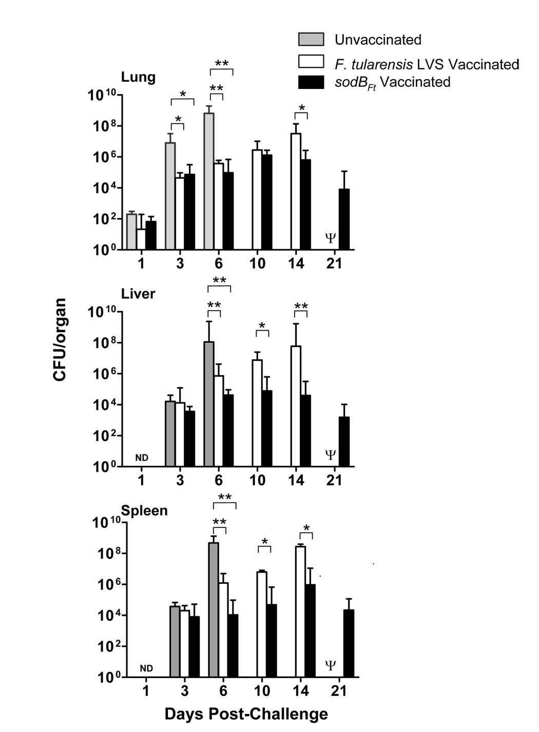
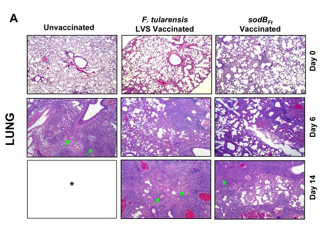
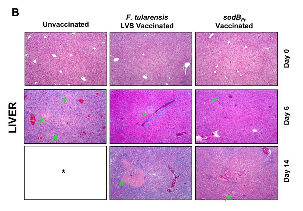
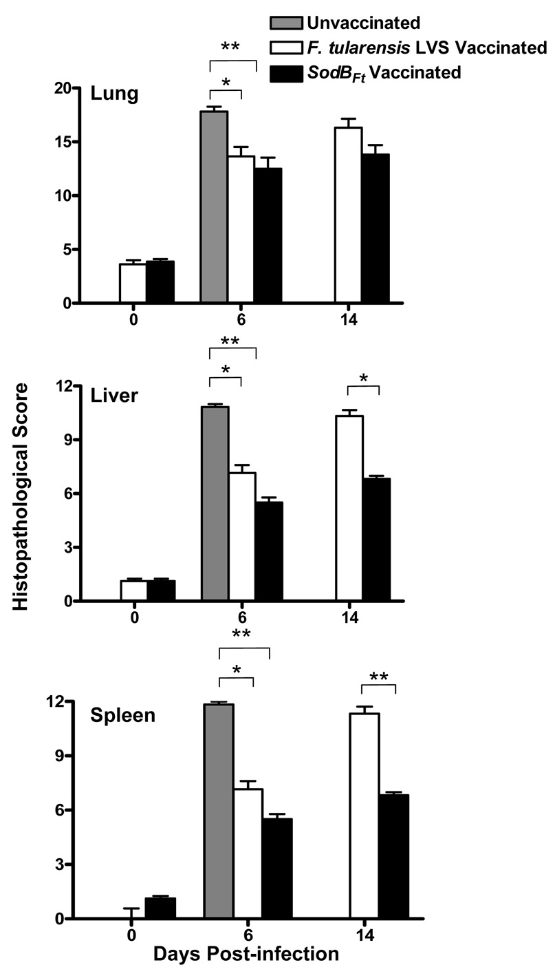
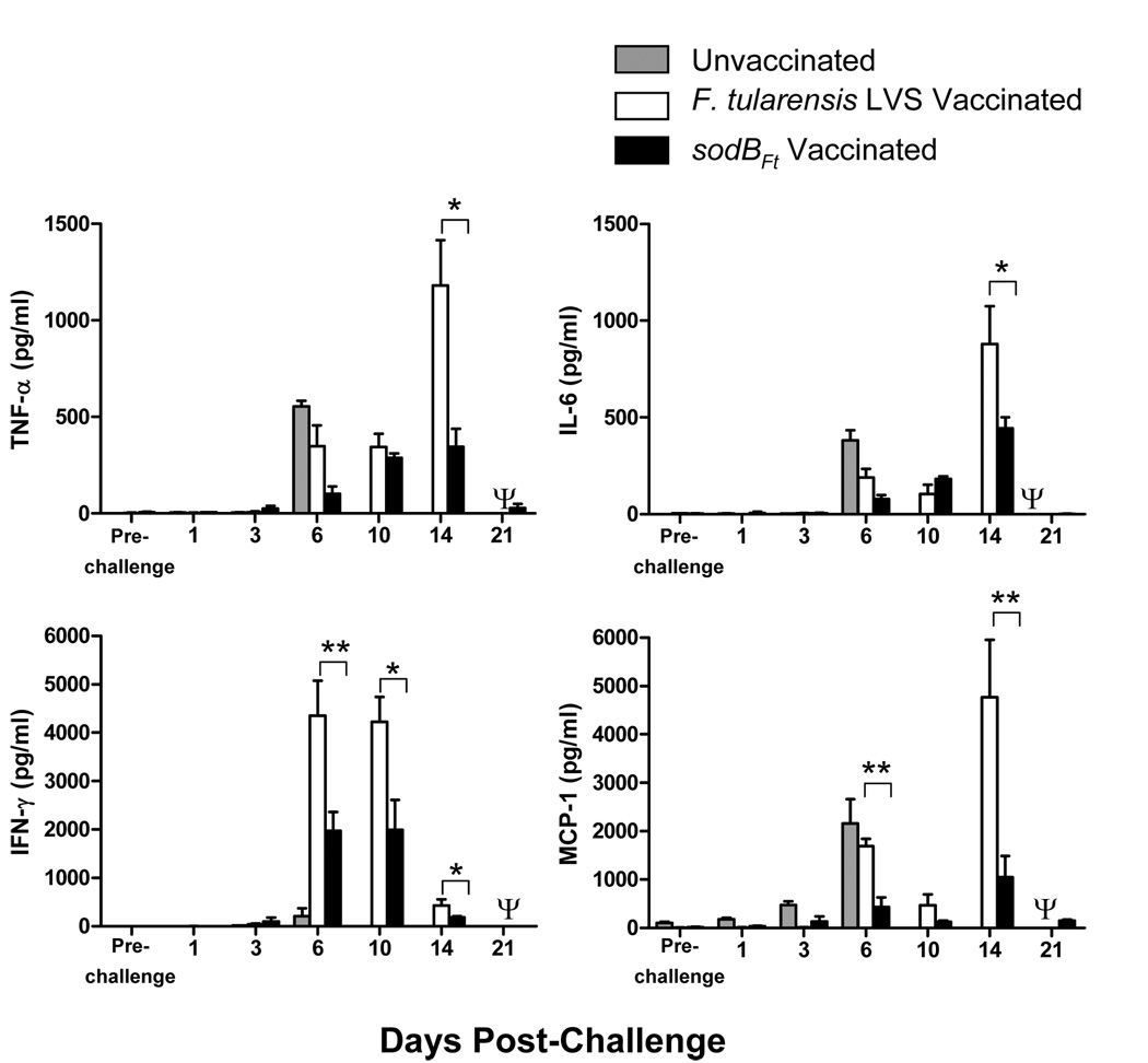
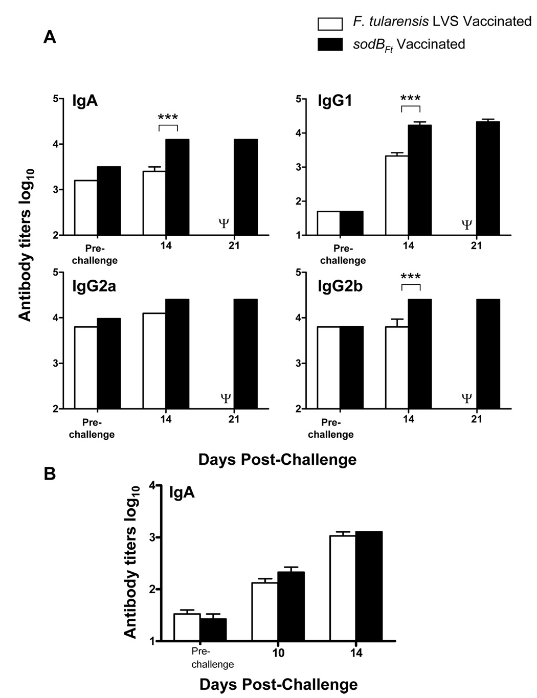
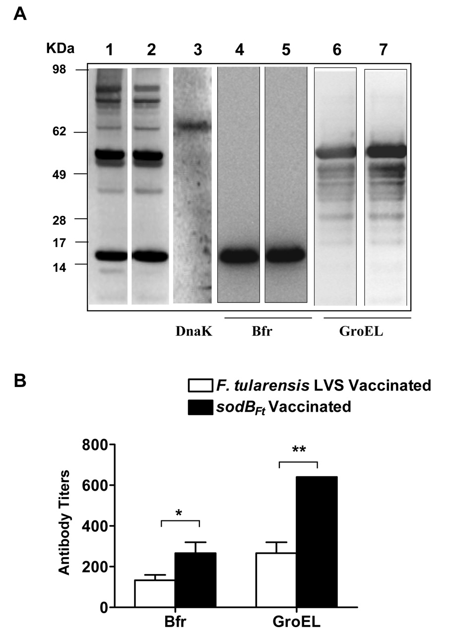
Vaccination of mice with Francisella tularensis live vaccine strain (LVS) mutants described so far have failed to induce protection in C57BL/6 mice against challenge with the virulent strain F. tularensis SchuS4. We have previously reported that a mutant of F. tularensis LVS deficient in iron superoxide dismutase (sodB(Ft)) is hypersensitive to oxidative stress and attenuated for virulence in mice. Herein, we evaluated the efficacy of this mutant as a vaccine candidate against respiratory tularemia caused by F. tularensis SchuS4. C57BL/6 mice were vaccinated intranasally (i.n.) with the sodB(Ft) mutant and challenged i.n. with lethal doses of F. tularensis SchuS4. The level of protection against SchuS4 challenge was higher in sodB(Ft) vaccinated group as compared to the LVS vaccinated mice. sodB(Ft) vaccinated mice following SchuS4 challenge exhibited significantly reduced bacterial burden in lungs, liver and spleen, regulated production of pro-inflammatory cytokines and less severe histopathological lesions compared to the LVS vaccinated mice. The sodB(Ft) vaccination induced a potent humoral immune response and protection against SchuS4 required both CD4 and CD8 T cells in the vaccinated mice. sodB(Ft) mutants revealed upregulated levels of chaperonine proteins DnaK, GroEL and Bfr that have been shown to be important for generation of a potent immune response against Francisella infection. Collectively, this study describes an improved live vaccine candidate against respiratory tularemia that has an attenuated virulence and enhanced protective efficacy than the LVS.


Figures

Figure 1
Figure 1. SodBFt vaccinated mice exhibit enhanced bacterial clearance following SchuS4 challenge
(A) C57BL/6 mice were vaccinated i.n. with F. tularensis LVS or sodBFt and challenged with F. tularensis SchuS4 on day 21 post-primary vaccination. At the times indicated, mice were sacrificed and homogenates of the lung, liver, and spleen were plated for determination of bacterial burden. Results shown are the mean ± SEM and are cumulative of two independent experiments conducted (n = 6–8 mice per group). * p < 0.05, ** p < 0.001 using the one-way ANOVA. Ψ All the LVS vaccinated mice died by day 15–17 post-challenge and hence were unavailable for comparisons.
Figure 2
Figure 2. SodBFt vaccinated mice exhibit less severe histopathological changes than LVS vaccinated mice following SchuS4 challenge
H and E stained sections from A. Lung; B. Liver and C. Spleen sections from unvaccinated and LVS or sodBFt vaccinated mice prior to challenge (day 0) and at days six and 14 post-SchuS4 challenge. Arrows in the lung panel indicate necrotizing pneumonia, in the liver indicate necrotic granulomas and those in the spleen indicate an area of intense lymphoproliferation and necrotizing splenitis. * All the unvaccinated mice died shortly after day 6 post-challenge. (Magnification × 40X).
Figure 3
Figure 3. Quantitation of histopathological lesions in the lung, liver and spleen from unvaccinated or vaccinated mice challenged with SchuS4
H and E stained tissue sections from unvaccinated and LVS or sodBFt vaccinated mice were evaluated for histopathological lesions prior to challenge (day 0) and at days six and 14 post SchuS4 challenge. The values represent cumulative histopathological scores (n=6 mice per group) based on the criteria described in Table 1. The results are expressed as mean ± SE and P values were determined using the Student’s t- test (*p < 0.05, **p < 0.01).
Figure 4
Figure 4. SodBFt vaccinated mice produce regulated levels of proinflammatory cytokines following SchuS4 challenge
Unvaccinated and SodBFt or LVS vaccinated and SchuS4 challenged C57BL/6 mice were sacrificed at the times indicated and cytokine levels were measured in homogenates of the lungs. The results shown are the mean ± SEM and are cumulative of two independent experiments conducted (n = 6–8 mice per time point). *p < 0.05, **p < 0.01 using the one-way ANOVA with Bonferroni’s multiple comparison test. Ψ All the LVS vaccinated mice died by day 15–17 post-challenge and hence were unavailable for comparisons.
Figure 5
Figure 5. SodBFt vaccinated C57BL/6 mice exhibit elevated levels of anti- F. tularensis specific antibodies following SchuS4 challenge
(A) Anti-F. tularensis antibody isotypes were determined by ELISA in sera, and (B) IgA levels in the lung homogenates from LVS or sodBFt vaccinated and SchuS4 challenged mice at the indicated times. The data represent an average of 3–4 mice per group. P values were determined using ANOVA. ** p < 0.01; *** p < 0.001. Ψ All the LVS vaccinated mice died by day 15–17 post-challenge and hence were unavailable for comparisons.
Figure 6
Figure 6. SodBFt exhibits increased levels of DnaK and GroEL as compared to F. tularensis LVS
(A) RT-PCR analysis to determine the transcript levels of DnaK and GroEL (B). qRT-PCR for the quantitation of DnaK and GroEL transcripts. The results are representative of three experiments conducted.
Figure 7
Figure 7. Determination of antibody responses against stress proteins GroEL, DnaK and Bfr in LVS or sodBFt vaccinated mice at day- 14 post-SchuS4 challenge
(A). The western blot analysis was performed as described in the materials and methods. The SchuS4 lysates were probed with pooled sera (n=3 mice) from LVS vaccinated mice (Lane 1), or sodBFt vaccinated mice (Lane 2) collected at day- 14 post-schuS4 challenge. The blots were stripped and reprobed with anti-DnaK antibodies to identify DnaK protein (Lane 3). For determination of antibody responses against Bfr and GroEL, purified recombinant proteins were probed with day- 14 post- SchuS4 challenge serum from LVS vaccinated (Lanes 4 and 6) and sodBFt vaccinated mice (Lanes 5 and 7), respectively. The protein bands observed for Bfr (Lanes 4–5), GroEL (Lane 6–7) and DnaK (Lane 3) corresponds to ~17 kDa, ~59 kDa and ~70 kDa bands in the SchuS4 lysates (Lane 1 and 2). (B). Quantitation of anti- Bfr and GroEL antibodies in day- 14 post- SchuS4 challenge serum from LVS and sodBFt vaccinated mice by ELISA. The data are expressed as mean ± S.D. (n=3 mice per group). P values were determined using Student’s t- test. * p ≤ 0.05; ** p < 0.01.

References

    1. Oyston PC, Sjostedt A, Titball RW. Tularaemia: bioterrorism defence renews interest in Francisella tularensis. Nat Rev Microbiol. 2004 Dec;2(12):967–978. - PubMed
    1. Saslaw S, Carlisle HN. Studies with tularemia vaccines in volunteers. IV. Brucella aggiutinins in vaccinated and nonvaccinated volunteers challenged with Pasteurella tularensis. Am J Med Sci. 1961 Aug;242:166–172. - PubMed
    1. Burke DS. Immunization against tularemia: analysis of the effectiveness of live Francisella tularensis vaccine in prevention of laboratory-acquired tularemia. J Infect Dis. 1977 Jan;135(1):55–60. - PubMed
    1. Hornick RB, Eigelsbach HT. Aerogenic immunization of man with live Tularemia vaccine. Microbiol Mol Biol Rev. 1966 Sep 1;30(3):532–538. - PMC - PubMed
    1. Eigelsbach HT, Hunter DH, Janssen WA, Dangerfield HG, Rabinowitz SG. Murine model for study of cell-mediated immunity: protection against death from fully virulent Francisella tularensis infection. Infect Immun. 1975 Nov;12(5):999–1005. - PMC - PubMed

Publication types

MeSH terms