. 2008 Aug 14;4(2):159-69.
doi: 10.1016/j.chom.2008.05.018.

Actin and intermediate filaments stabilize the Chlamydia trachomatis vacuole by forming dynamic structural scaffolds

Affiliations

Actin and intermediate filaments stabilize the Chlamydia trachomatis vacuole by forming dynamic structural scaffolds

Yadunanda Kumar et al. Cell Host Microbe. .

Abstract

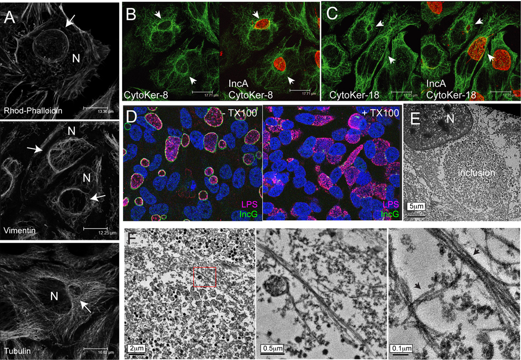
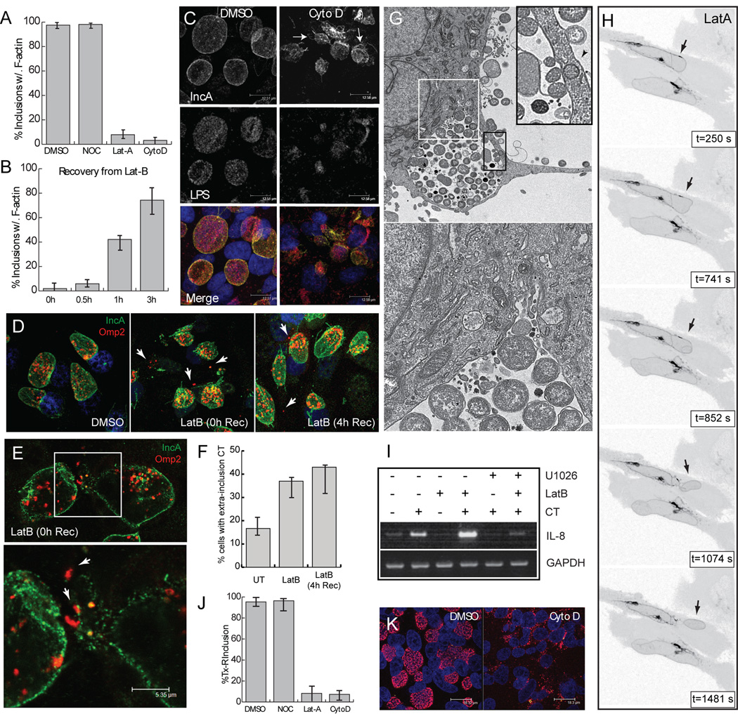
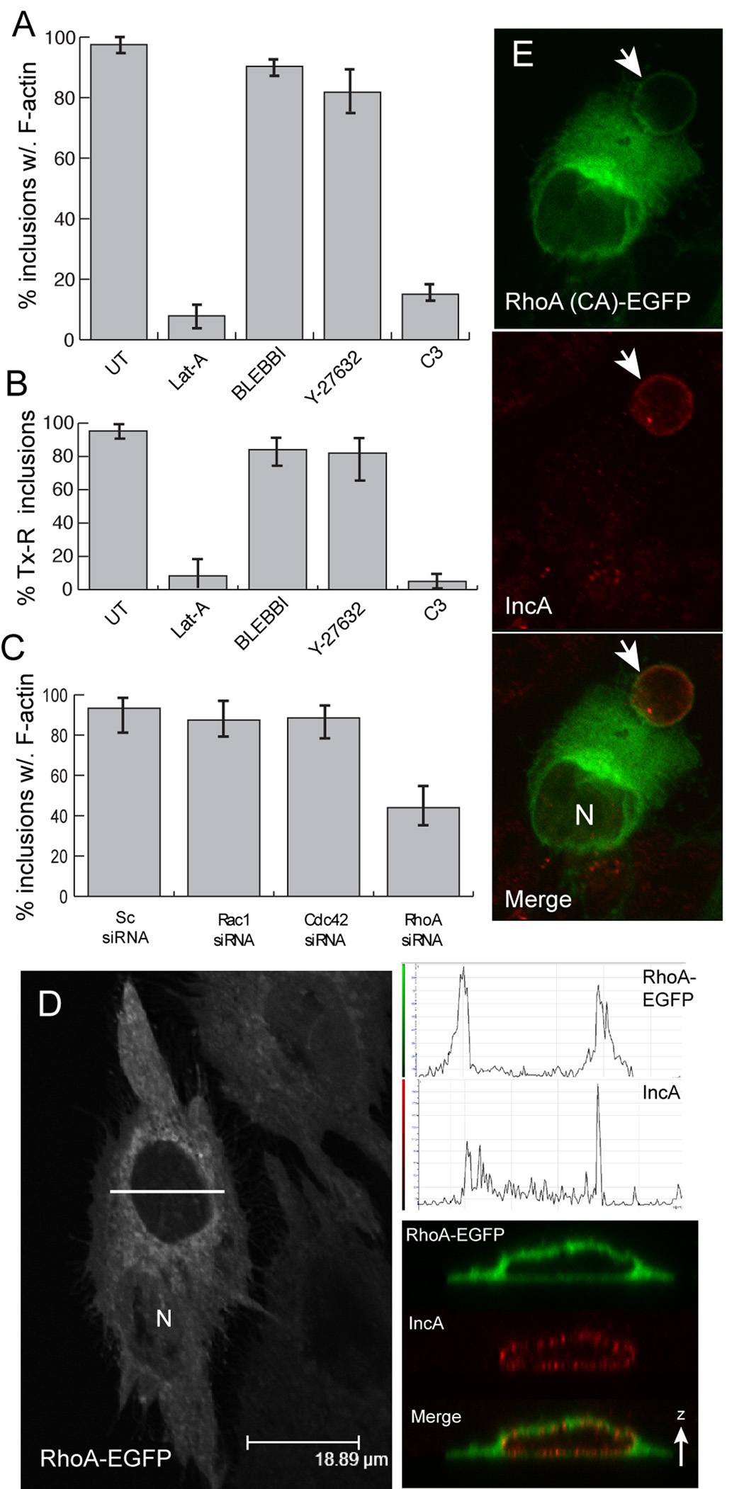
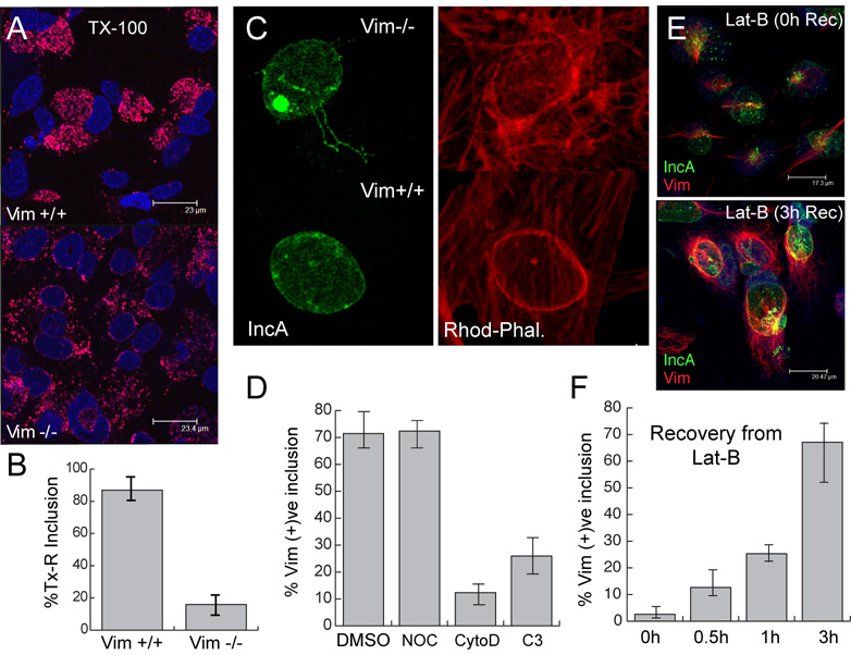
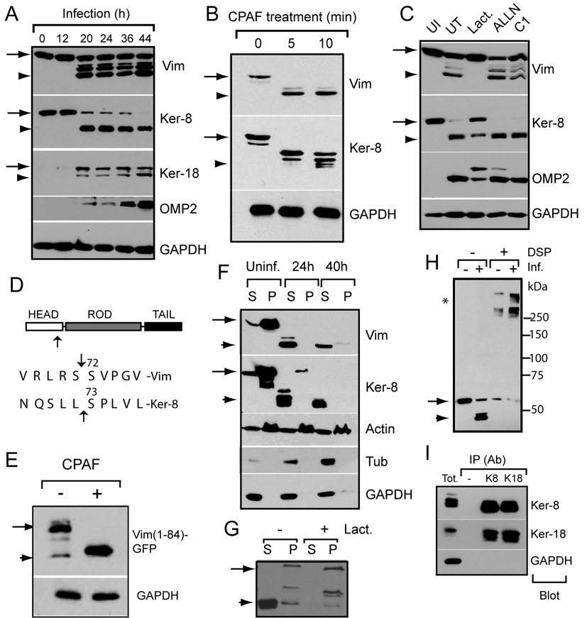
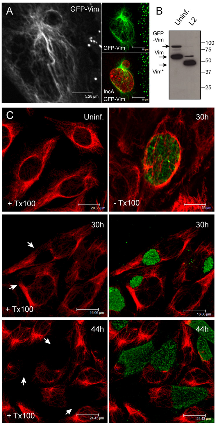
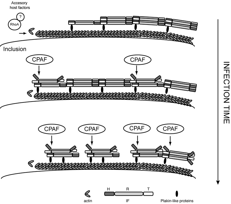
The obligate intracellular bacterial pathogen Chlamydia trachomatis replicates within a large vacuole or "inclusion" that expands as bacteria multiply but is maintained as an intact organelle. Here, we report that the inclusion is encased in a scaffold of host cytoskeletal structures made up of a network of F-actin and intermediate filaments (IF) that act cooperatively to stabilize the pathogen-containing vacuole. Formation of F-actin at the inclusion was dependent on RhoA, and its disruption led to the disassembly of IFs, loss of inclusion integrity, and leakage of inclusion contents into the host cytoplasm. In addition, IF proteins were processed by the secreted chlamydial protease CPAF to form filamentous structures at the inclusion surface with altered structural properties. We propose that Chlamydia has co-opted the function of F-actin and IFs to stabilize the inclusion with a dynamic, structural scaffold while minimizing the exposure of inclusion contents to cytoplasmic innate immune-surveillance pathways.


Figures

Figure 1
Figure 1. Actin and intermediate filaments are reorganized at the periphery of the C.trachomatis inclusion
A. Architecture of the host cytoskeleton in Chlamydia-infected cells. HeLa cells were infected with C. trachomatis LGV-L2 for 26–30h, fixed and processed for immunofluorescence microscopy. F-actin was detected with rhodamine-phalloidin, and intermediate filaments (IF) and microtubules were detected with antibodies to vimentin, α-tubulin, respectively. Note the assembly of actin and vimentin filaments at the inclusion (arrows). B–C. Cages of cytokeratin-8 and -18 filaments assemble at the inclusion. HeLa cells were infected as in A, fixed and cytokeratins detected with antibodies to cytokeratin-8 (B) and -18 (C). Inclusions (arrows) were detected with anti-IncA antibodies (red). D–E. The lumenal contents of inclusions are resistant to detergent extraction. HeLa cells were infected for 30h and either left untreated (left) or treated (right) with 1% Triton X-100 (Tx-100) for 5min at 4°C. Cells were fixed and stained with anti chlamydial LPS (red) and IncG (green) antibodies, and the DNA stain TO-PRO-3 (blue). Transmission electron micrographs of detergent extracted cells show the intact morphology of inclusion and the loss of membranes at the periphery of the inclusion (E). F. Parallel filaments localize to the inclusion periphery. Transmission electron micrographs reveal F-actin (~10nm) filament bundles (arrows) associated with the periphery of the inclusion.
Figure 2
Figure 2. F-actin assembly is required for inclusion integrity and stability
A–B. F-actin recruitment to the inclusion is dynamic. HeLa cells were infected with LGV-L2 for 30h and the number of F-actin positive inclusions was quantified after treatment with latrunculin A (Lat-A 400nM, 15min), nocodazole (Noc, 10µM, 2h) and cytochalasin D (Cyto D 1µM, 30min) (A) or after recovery from a 30min treatment with latrunculin B (Lat-B, 0.5 µM) (B) (n=150). C. Disruption of F-actin leads to alterations in inclusion membrane morphology. HeLa cells infected with LGV-L2 for 30h were treated with DMSO or Cyto D (1µM, 30min) and immunostained with anti-IncA and chlamydial LPS antibodies to detect inclusion membranes and bacteria, respectively. Note the accumulation of deformed inclusions (arrows). D–G. Disruption of F-actin leads to the release of inclusion contents into the cytoplasm. Infected HeLa cells were treated with DMSO or Lat-B, followed by washout of the drug for 4h. Cells were permeabilized with digitonin and IncA and Omp2 detected by immunofluorescence microscopy. Note the presence of Omp2-positive bacteria (arrows) not associated with IncA (D–E). Electron micrographs of LatA-treated cells (G) indicate potential rupture sites at the inclusion membrane (white box bottom panel) and bacteria (arrow) in the cytoplasm (black box upper right). H. Fragmentation of inclusions in response to LatA treatment. Time lapse series of infected cells where inclusion membranes are labeled with Lda3-EGFP. Note formation of satellite inclusions (arrows) after Lat-treatment. I. F-actin disruption leads to increased IL-8 activation in infected cells. IL-8 expression levels were assessed by RT-PCR of total RNA isolated from infected and uninfected HeLa cells after Lat-B treatment (as in D). The ERK inhibitor U1026 was added as a control for Chlamydia-mediated IL-8 activation. J–K. Disruption of F-actin renders the inclusion sensitive to detergent extraction. Infected HeLa (30h) were treated as in A. The percentage of Tx-100-resistant inclusions (Fig. 1D), as assessed by staining with anti-chlamydial LPS (G) was quantified (n=200). The error bars represent positive and negative deviations from the mean of three independent experiments.
Figure 3
Figure 3. F-actin recruitment to the inclusion requires RhoA function
A–C. RhoA is required for F-actin assembly and inclusion stability. HeLa cells were infected with LGV-L2 for 30h and treated with Lat-A, Blebbistatin (50µM, 1h), the ROCK inhibitor Y-27632 (20µM, 1h) or C3-transferase (3µg/ml, 6h). Cells were fixed and stained for F-actin and IncA or extracted with 1% Tx-100 prior to fixation followed by immunostaining with anti-LPS antibodies. The percentage of inclusions with intact F-actin rings (A) or that were resistant to detergent extraction (B) was determined as in Fig. 2. UT: Untreated. C. The expression of Rho proteins was inhibited by transfection of specific siRNAs and the percentage of inclusions with F-actin-ring determined as in Fig. 2 (n=150). A scrambled (Sc) siRNA (Qiagen) were used as a non-specific control. D–E. RhoA is recruited to the periphery of the inclusion. HeLa cells were infected with RhoA-EGFP (D) or a constitutively active RhoA variant Q67L (E). Note the accumulation of the tagged proteins at the periphery of the IncA-positive inclusion membranes. Corresponding zy confocal scans and quantification of EGFP and IncA signal intensity at a representative cross-section of the inclusion is shown (D right panels).
Figure 4
Figure 4. Vimentin filaments contribute to inclusion stability
A–B. Inclusions in vimentin deficient fibroblasts are sensitive to detergent extraction. Mouse embryo fibroblasts (MEF) derived from vimentin knockout mice (MTF16 -Vim−/−) or wild-type littermate controls (MTF6 -Vim+/+) were infected with LGV-L2 for 30h, treated live with 1% Tx-100, processed for immunofluorescence as in Fig. 1D (A), and the frequency of detergent resistant inclusions assessed (B). C. Morphology of actin rings in vimentin deficient MEFs. Vim−/− and Vim+/+ MEFs were infected with LGV-L2 for 30h and F-actin detected with Rhodamine-phalloidin. Note diffuse actin ring surrounding IncA positive inclusion in Vim−/− MEFs. D–F. Vimentin recruitment to the inclusion requires Rho-dependent F-actin assembly. HeLa cells infected with LGV-L2 for 30h were treated with Noc, CytoD or C3 as in Fig 2, fixed and immunostained for vimentin and IncA. The percentage of vimentin-positive inclusions was determined in the presence of inhibitors (D) or after removal of Lat-B for 0–3 h (F) (n=150). Note reassembly of cages of vimentin (red) filaments around inclusions (arrows) after removal of Lat B (E). Host and bacterial DNA was detected with TO-PRO-3 (blue)
Figure 5
Figure 5. Proteolytic processing of IF proteins by the chlamydial proteasome like activity factor (CPAF) alters its cytoskeletal properties
A–D. The Head domain of IFs is cleaved by CPAF during infection. HeLa cells were infected with LGV-L2 for the indicated times. Total cell lysates were prepared and proteins were identified by SDS-PAGE followed by immunoblots with specific antibodies. Note processing of vimentin, cytokeratin-8 and -18 (A). CPAF cleaved IFs in vitro (see methods) to generate fragments similar to those observed in vivo (B). Cleavage was inhibited in vivo by pretreatment with lactacystin but not by proteasome (ALLN) or Type III secretion inhibitors (C1) (C). The CPAF cleavage site was determined by amino-terminal sequencing vimentin purified from infected cells and shown to map to Ser72 in the Head domain of the IF protein (D). For comparison, the CPAF cleavage site for keratin-8 is shown (Dong et al., 2004). E. The Rod and Tail domains are not required for CPAF recognition of vimentin. Cell lysates from HeLa cells expressing the Head domain of vimentin (aa 1–84) fused to GFP were treated with CPAF for 30min and analyzed by immunoblots. F–G. The cytoskeletal properties of IFs are altered by CPAF. HeLa cells were infected with LGV-L2 for 0, 24h or 40h, extracted with 1% Tx X-100 and subjected to differential centrifugation. Proteins in the detergent soluble and insoluble fractions were identified by immunobloting with specific antibodies. Note partitioning of the IF cleavage products (arrowheads) with the detergent soluble fractions. Lactacystin treatment prevented the partitioning of IFs to soluble fractions in infected cells (G). H. Cleaved vimentin is incorporated into high molecular weight complexes. Infected and uninfected HeLa cells were treated with the crosslinker DSP and the vimentin cross-linked complexes (*) were identified in non-reduced samples by immunoblotting. I. The formation of keratins 8/18 heterodimers is resistant to CPAF-mediated cleavage. Keratin-8 and keratin-18 were immunoprecipitated (IP) from infected HeLa lysates and the co-IP of their cognate filament partners was assessed by immunoblots. Arrows indicate full length IF protein and arrowheads indicate processed forms of IF proteins.
Figure 6
Figure 6. Vimentin filaments at the periphery of the inclusion are sensitive to detergent extraction
A–B. The cleaved Head domain of vimentin remains associated with inclusion filaments. HeLa cells expressing vimentin with an amino terminal GFP fusion (GFP-Vim) were infected with LGV-L2 for 24h and analyzed by fluorescence microscopy (A). Note GFP-vimentin in inclusion associated filaments (A) despite complete cleavage of the tagged protein (B). C. Vimentin filaments at the periphery of the inclusion are preferentially sensitive to detergent extraction. HeLa cells were infected with LGV-L2 for 30–44h, solubilized with Tx-100 and immunostained with anti-vimentin (red) and anti-chlamydial (green) antibodies. Note progressive detergent sensitivity of vimentin filaments associated with the inclusion periphery. As a reference note compact IF cage present in non detergent-treated infected cells (upper right panel).
Figure 7
Figure 7. A model for actin and intermediate filament assembly at the surface of the Chlamydia inclusion
C. trachomatis effectors at the inclusion membrane recruit RhoA to trigger F-actin assembly. F-actin at the inclusion helps recruit and stabilize IFs possibly via ‘linker’ molecules to form a stable support cage for the inclusion. As the inclusion expands the secreted chlamydial protease-CPAF progressively cleaves the Head domain of preassembled filaments to increase their flexibility while retaining some of their structural functions.

Comment in

Similar articles

Cited by

References

    1. Aktories K, Just I. Clostridial Rho-inhibiting protein toxins. Curr Top Microbiol Immunol. 2005;291:113–145. - PubMed
    1. Anes E, Kuhnel MP, Bos E, Moniz-Pereira J, Habermann A, Griffiths G. Selected lipids activate phagosome actin assembly and maturation resulting in killing of pathogenic mycobacteria. Nat Cell Biol. 2003;5:793–802. - PubMed
    1. Batchelor M, Guignot J, Patel A, Cummings N, Cleary J, Knutton S, Holden DW, Connerton I, Frankel G. Involvement of the intermediate filament protein cytokeratin-18 in actin pedestal formation during EPEC infection. EMBO Rep. 2004;5:104–110. - PMC - PubMed
    1. Beatty WL. Trafficking from CD63-positive late endocytic multivesicular bodies is essential for intracellular development of Chlamydia trachomatis. J Cell Sci. 2006;119:350–359. - PubMed
    1. Belland R, Ojcius DM, Byrne GI. Chlamydia. Nat Rev Microbiol. 2004;2:530–531. - PubMed

Publication types

MeSH terms