Review
. 2008 Sep;30(9):811-21.
doi: 10.1002/bies.20804.

Microfluidics meet cell biology: bridging the gap by validation and application of microscale techniques for cell biological assays

Affiliations
Review

Microfluidics meet cell biology: bridging the gap by validation and application of microscale techniques for cell biological assays

Amy L Paguirigan et al. Bioessays. 2008 Sep.

Abstract

Microscale techniques have been applied to biological assays for nearly two decades, but haven't been widely integrated as common tools in biological laboratories. The significant differences between several physical phenomena at the microscale versus the macroscale have been exploited to provide a variety of new types of assays (such as gradient production or spatial cell patterning). However, the use of these devices by biologists seems to be limited by issues regarding biological validation, ease of use, and the limited available readouts for assays done using microtechnology. Critical validation work has been done recently that highlights the current challenges for microfluidic methods and suggest ways in which future devices might be improved to better integrate with biological assays. With more validation and improved designs, microscale techniques hold immense promise as a platform to study aspects of cell biology that are not possible using current macroscale techniques.


Figures

Figure 1
Figure 1
Examples of microfluidic gradient production devices. Flow based gradients like that shown in (a) are based on diffusional mixing solely at the interface between fluid streams. Here two solutions with different concentrations of the solute of interest (0% and 100% of the desired final concentration in this case) are introduced to the inputs of a gradient generation network. Diffusional mixing occurs at the interfaces of the fluid streams and creates a gradient of a defined profile (dependent on input concentrations) at the point labeled Migration channel where cells are treated with the flowing gradient of interest. Static gradient systems like that shown in (b) can be used to create stable gradients in a static fluid, by addition of fluid of the maximum concentration at the Source and allowing the solute to diffuse to the sink, thus exposing cells in the channel to a gradient of the factor. Adapted from: (a) Biomedical Microdevices, A parallel-gradient microfluidic chamber for quantitative analysis of breast cancer cell chemotaxis, 8, 2006, page 109–118, Saadi W, Wang SJ, Lin F, Jeon NL, Figure 1 with kind permission from Springer Science +Business Media: and (b) from Abhyankar VV, Lokuta MA, Huttenlocher A, Beebe DJ. 2006. Characterization of a membrane-based gradient generator for use in cell-signaling studies. Lab Chip 6:389393. Reproduced by permission of The Royal Society of Chemistry, http://dx.doi.org/10.1039/b514133h.
Figure 2
Figure 2
A comparison between volume densities of culture conditions in traditional, macroscale culture in 6-well plates and in microscale, microchannel culture. Given the same cell surface density, even a rather large microchannel (750 μm wide, 5 mm long, and 250 μm tall) can provide a volume density 2–4 times that of a traditional well in a 6-well plate, use 250 times fewer cells, and 500–1000 times less media and costly reagents.
Figure 3
Figure 3
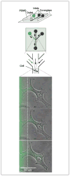
Illustration of a microfluidic device capable of treating subcellular domains with specific reagents while leaving the rest of a cell or region unaffected. Schematics of the device are shown at the top in which the green channels represent the channel in which dye is included. Fluid from the three inputs flows alongside one another and only mix via diffusion, allowing part of the cell shown below to be stained prior to significant mixing of the reagent. In this case, BCE cells are shown in the lower panels after being labeled with MitoTracker Green for 5, 11 and 35 minutes of exposure to the dye, from top to bottom respectively. Reprinted from Chemistry and Biology, 10/2, Takayama S, Ostuni E, LeDuc P, Naruse K, Ingber DE, Whitesides GM, Selective chemical treatment of cellular microdomains using multiple laminar streams., 123–130, Copyright 2003, with permission from Elsevier.
Figure 4
Figure 4
Microfluidic channels fabricated from poly(dimethyl siloxane), (PDMS) have been shown to absorb small hydrophobic molecules. a: Quinine (fluoresceces at pH2, but not at pH7) was put into a channel and then washed out with pH2 water and fluorescence images of the channel taken. b: If quinine is incubated for 5 minutes in pH7 water in the channel no fluorescence is seen, but after the channel is washed with pH2 water, quinine begins to leach back into solution from the PDMS channel walls and remains until it is washed again. c: A similar phenomenon was shown for Nile Red, as even after the channel is washed with detergent and water, significant fluorescence indicates that the Nile Red was absorbed into the walls of the channels. Adapted from Toepke MW, Beebe DJ. 2006. PDMS absorption of small molecules and consequences in microfluidic applications. Lab On A Chip 6:1484–1486. Reproduced by permission of The Royal Society of Chemistry http://dx.doi.org/10.1039/b612140c.

Comment in

References

    1. Sims CE, Allbritton NL. Analysis of single mammalian cells on-chip. Lab On A Chip. 2007;7:423–440. - PubMed
    1. Keenan TM, Folch A. Biomolecular gradients in cell culture systems. Lab Chip. 2008;8:34–57. Epub 2007 Dec 6. - PMC - PubMed
    1. Meyvantsson I, Warrick JW, Hayes S, Skoien A, Beebe DJ. Automated cell culture in high density tubeless microfluidic device arrays. Lab Chip. 2008;8(5):717–24. - PubMed
    1. Tan W, Desai TA. Microfluidic patterning of cells in extracellular matrix biopolymers: effects of \ channel size, cell type, and matrix composition on pattern integrity. Tissue Eng. 2003;9:255–567. - PubMed
    1. Fisher RJ, Peattie RA. Controlling tissue microenvironments: biomimetics, transport phenomena, and reacting systems. Adv Biochem Eng Biotechnol. 2007;103:1–73. - PubMed

Publication types

MeSH terms