

Computerized pathological image analysis for neuroblastoma prognosis
- PMID: 18693847
- PMCID: PMC2655895
Computerized pathological image analysis for neuroblastoma prognosis
Abstract
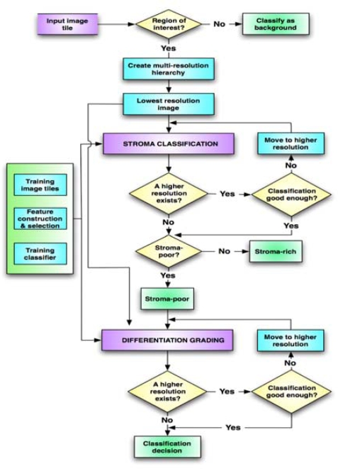
We present a pathological image analysis system for the computer-aided prognosis of neuroblastoma, a childhood cancer. The image analysis system automatically classifies Schwannian stromal development of pathological tissues and determines the grade of differentiation. Due to the demanding computational cost of processing large digitized slides, the system was implemented on a cluster of computers with automated load balancing within a multi-resolution framework. In our experiments, the overall accuracies for stromal classification and the grade of differentiation were 96.6% and 95.3%, respectively. Additionally, the multi-resolution framework reduced the run time of the single resolution approach by 53% and 34% on average for stromal classification and grade of differentiation, respectively. For these two cases, parallelization on a 16-node cluster reduced the sequential run time by 92% and 88% on average. Accuracy and efficiency of these techniques are promising for the development a computer-assisted neuroblastoma prognosis system.
Figures
References
-
- Shimada H, Chatten J, Newton WA, Jr, Sachs N, Hamoudi AB, Chiba T, Marsden HB, Misugi K. “Histopathologic prognostic factors in neuroblastic tumors: definition of subtypes of ganglioneuroblastoma and an age-linked classification of neuroblastomas,”. J Natl Cancer Inst. 1984 Aug;73:405–16. - PubMed
-
- Shimada H, Ambros IM, Dehner LP, Hata J, Joshi VV, Roald B, Stram DO, Gerbing RB, Lukens JN, Matthay KK, Castleberry RP. “The International Neuroblastoma Pathology Classification (the Shimada system),”. Cancer. 1999 Jul 15;86:364–72. - PubMed
-
- Ojala T, Pietikainen M, Maenpaa T. “Multiresolution Gray-Scale and Rotation Invariant Texture Classification with Local Binary Patterns,”. IEEE Trans. on PAMI. 2002;24:17.
-
- Tuceryan M, Jain AK. The Handbook of Pattern Recognition and Computer Vision (2nd Edition) 2 ed. World Scientific Publishing Co; 1998. “Texture Analysis,”; p. 41.
-
- Adelson EH, Anderson CH, Bergen JR, Burt PJ, Ogden J. “Pyramid methods in image processing,”. RCA Engineer. 1984;29:9.
Publication types
MeSH terms
Grants and funding
LinkOut - more resources
Full Text Sources
Other Literature Sources
Medical