Review
. 2009 Feb;15(2):205-19.
doi: 10.1089/ten.tea.2008.0131.

Engineered microenvironments for controlled stem cell differentiation

Affiliations
Review

Engineered microenvironments for controlled stem cell differentiation

Jason A Burdick et al. Tissue Eng Part A. 2009 Feb.

Abstract

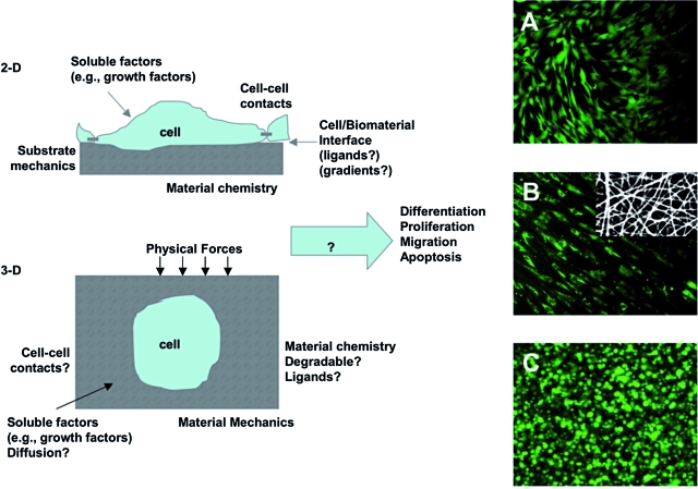
In a developing organism, tissues emerge from coordinated sequences of cell renewal, differentiation, and assembly that are orchestrated by spatial and temporal gradients of multiple regulatory factors. The composition, architecture, signaling, and biomechanics of the cellular microenvironment act in concert to provide the necessary cues regulating cell function in the developing and adult organism. With recent major advances in stem cell biology, tissue engineering is becoming increasingly oriented toward biologically inspired in vitro cellular microenvironments designed to guide stem cell growth, differentiation, and functional assembly. The premise is that to unlock the full potential of stem cells, at least some aspects of the dynamic three-dimensional (3D) environments that are associated with their renewal, differentiation, and assembly in native tissues need to be reconstructed. In the general context of tissue engineering, we discuss the environments for guiding stem cell function by an interactive use of biomaterial scaffolds and bioreactors, and focus on the interplay between molecular and physical regulatory factors. We highlight some illustrative examples of controllable cell environments developed through the interaction of stem cell biology and tissue engineering at multiple levels.


Figures

FIG. 1.
FIG. 1.
Manipulating the stem cell microenvironment in 2D and 3D. Schematic of controllable parameters (e.g., matrix properties and culture environment) for altering stem cell microenvironmental behavior in both 2D and 3D. Human MSC morphology (stained with Live/Dead) when seeded in 2D on a biodegradable elastomer (A), on the surface of an electrospun fibrous scaffold from a biodegradable elastomer and gelatin (autofluoresces red) composite (B, inset is SEM of scaffold), and when encapsulated in 3D in a photocrosslinked hyaluronic acid hydrogel (C). These are examples where the biomaterial structure dictates the cellular morphology. Work by the Burdick laboratory and previously unpublished. Color images available online at www.liebertonline.com/ten.
FIG. 2.
FIG. 2.
Matrix chemistry controls stem cell differentiation. Colonies of human ESCs encapsulated in 3D hyaluronic acid hydrogels and stained for markers of undifferentiation (green, Oct4; red, SSEA4 (A) and compared using light microscopy (left) and histology (right) to human ESCs encapsulated in dextran hydrogels (B). Colonies of hESCs maintain undifferentiated in HA hydrogels and spontaneously differentiated in dextran hydrogels with a similar chemical structure, indicating the importance of biomaterial chemistry on cellular interactions. Reprinted with permission from Gerecht et al. Copyright (2007) National Academy of Sciences, USA. Color images available online at www.liebertonline.com/ten.
FIG. 3.
FIG. 3.
Matrix mechanics directs stem cell differentiation. Matrix mechanics–dependent differentiation of human MSCs stained for β3-Tubulin, MyoD, and CBFα1 as markers of neurogenic, myogenic, and osteogenic differentiation, respectively (scale bar = 5 μm). MSC differentiation correlates to tissue-specific mechanical properties (e.g., soft leads to neural differentiation, whereas stiff leads to osteogenic differentiation). Reprinted with permission from Engler et al. Color images available online at www.liebertonline.com/ten.
FIG. 4.
FIG. 4.
Cell spreading controls stem cell lineage specification. Human MSC adipogenic (A, Oil Red O stain) and osteogenic (B, Alkaline Phosphatase stain) differentiation in response to cell spreading through size of adhesive islands. Quantification indicates that adipogenic differentiation is favored on smaller islands, whereas osteogenic is enhanced on larger islands where more spreading is allowed (C). Reprinted with permission from McBeath et al. Color images available online at www.liebertonline.com/ten.
FIG. 5.
FIG. 5.
Perfusion bioreactors for enhanced mass transport. Left: example of an integrated perfusion bioreactor system for cell seeding and prolonged culture of tissue constructs within a single device. (A) Cell seeding pathway: alternating bi-directional flow of cell suspension, without cell recirculation through the pump. (B) Cultivation pathway: transfer from seeding to cultivation is made by simply diverting the medium flow to a separate perfusion loop for prolonged culture. (C) Scaffold chamber: the scaffold is inserted into a removable holder and held in place by its outer 1 mm periphery. A straight region helps to fully develop the flow before reaching the construct. Right: Simple bioreactor system for tissue engineering of bone and osteochondral grafts that enables cultivation of up to six tissue constructs simultaneously, with direct perfusion and imaging capability. (D) Schematic presentation of the bioreactor system. The flow channels in the bioreactor provide an even distribution of medium flow between the six constructs (4–10 mm in diameter, up to 7 mm high) that are press-fit into the culture wells. (E) An example of a cartilage-bone plug from a gel layer overlaying the porous scaffold; both phases are seeded with human MSCs, and the construct is cultured in the perfusion bioreactor (D). (F) Example of an anatomically correct construct of a human temporomandibular joint condyle that was cultured in a bioreactor (D). Images (A)–(C) are reproduced with permission from Wendt et al. Color images available online at www.liebertonline.com/ten.
FIG. 6.
FIG. 6.
Mechanical stimulation bioreactors for functional tissue engineering. (A) Bioreactor system for dynamic deformational loading of engineered cartilage. Constructs are placed in the base of a standard Petri dish modified with a custom agarose template to maintain positioning during loading, which is carried out by applying sinusoidal deformation. Reproduced with permission from Hung et al. (B) Bioreactor system with pulsatile flow for engineering blood vessels. Fibrous tubular scaffolds seeded with aortic smooth muscle cells were placed over silicone tubing and subjected to pulsatile flow inducing 5% radial strain. After 8 weeks of culture, the silicone tubing was removed and a confluent layer of endothelial cells was formed in vessel lumens. Reproduced with permission from Niklason et al. (C) Bioreactor with mechanical stimulation for cardiac tissue engineering. Top left: Casting mold. Top right: Cells seeded into a collagen gel and cultured without stimulation for 1–4 days. Bottom left: Stretch apparatus for the application of unidirectional and cyclic stretch (10%, 2 Hz). Bottom right: Multiple rings connected into a construct for implantation studies. Reproduced with permission from Zimmermann et al. Color images available online at www.liebertonline.com/ten.
FIG. 6.
FIG. 6.
Mechanical stimulation bioreactors for functional tissue engineering. (A) Bioreactor system for dynamic deformational loading of engineered cartilage. Constructs are placed in the base of a standard Petri dish modified with a custom agarose template to maintain positioning during loading, which is carried out by applying sinusoidal deformation. Reproduced with permission from Hung et al. (B) Bioreactor system with pulsatile flow for engineering blood vessels. Fibrous tubular scaffolds seeded with aortic smooth muscle cells were placed over silicone tubing and subjected to pulsatile flow inducing 5% radial strain. After 8 weeks of culture, the silicone tubing was removed and a confluent layer of endothelial cells was formed in vessel lumens. Reproduced with permission from Niklason et al. (C) Bioreactor with mechanical stimulation for cardiac tissue engineering. Top left: Casting mold. Top right: Cells seeded into a collagen gel and cultured without stimulation for 1–4 days. Bottom left: Stretch apparatus for the application of unidirectional and cyclic stretch (10%, 2 Hz). Bottom right: Multiple rings connected into a construct for implantation studies. Reproduced with permission from Zimmermann et al. Color images available online at www.liebertonline.com/ten.
FIG. 7.
FIG. 7.
Microarray bioreactors for in vitro studies. (A) The micro-bioreactor wells (3.5 mm in diameter) are arranged in an array. Each of three inlets delivers medium (red) through the flow transducers to four wells (orange) via microfluidic channels (100 μm wide); waste medium exits each bioreactor via a separate set of channels (yellow). (B) Two configurations were used: a bottom inlet/outlet (BIO) configuration, and a middle inlet/outlet (MIO) configuration (right) that allows for 3D cultivation. (C) Image of a single MBA with compression frame and fluidic connections. (D) Experimental setup. MBAs and medium collectors are placed in an incubator, and the medium reservoirs are maintained external to the incubator in an ice bath. (E) Schematic presentation of the human ESC culture in the microarray bioreactor system. Images (A)–(D) are from Figallo et al. and reproduced by permission of The Royal Society of Chemistry. Color images available online at www.liebertonline.com/ten.

References

    1. Harrison R. Observations on the living developing nerve fiber. Anat Rec. 1907;1:116–118.
    1. Jacks T. Weinberg R.A. Taking the study of cancer cell survival to a new dimension. Cell. 2002;111:923–925. - PubMed
    1. Abbott A. Cell culture: biology's new dimension. Nature. 2003;424:870. - PubMed
    1. Weaver V.M. Petersen O.W. Wang F. Larabell C.A. Briand P. Damsky C. Bissell M.J. Reversion of the malignant phenotype of human breast cells in three-dimensional culture and in vivo by integrin blocking antibodies. J Cell Biol. 1997;137:231–245. - PMC - PubMed
    1. Griffith L.G. Swartz M.A. Capturing complex 3D tissue physiology in vitro. Nat Rev Mol Cell Biol. 2006;7:211–224. - PubMed

Publication types

Substances