. 2008 Aug 11:8:229.
doi: 10.1186/1471-2407-8-229.

Cigarette smoke induces endoplasmic reticulum stress and the unfolded protein response in normal and malignant human lung cells

Affiliations

Cigarette smoke induces endoplasmic reticulum stress and the unfolded protein response in normal and malignant human lung cells

Ellen Jorgensen et al. BMC Cancer. .

Abstract

Background: Although lung cancer is among the few malignancies for which we know the primary etiological agent (i.e., cigarette smoke), a precise understanding of the temporal sequence of events that drive tumor progression remains elusive. In addition to finding that cigarette smoke (CS) impacts the functioning of key pathways with significant roles in redox homeostasis, xenobiotic detoxification, cell cycle control, and endoplasmic reticulum (ER) functioning, our data highlighted a defensive role for the unfolded protein response (UPR) program. The UPR promotes cell survival by reducing the accumulation of aberrantly folded proteins through translation arrest, production of chaperone proteins, and increased degradation. Importance of the UPR in maintaining tissue health is evidenced by the fact that a chronic increase in defective protein structures plays a pathogenic role in diabetes, cardiovascular disease, Alzheimer's and Parkinson's syndromes, and cancer.

Methods: Gene and protein expression changes in CS exposed human cell cultures were monitored by high-density microarrays and Western blot analysis. Tissue arrays containing samples from 110 lung cancers were probed with antibodies to proteins of interest using immunohistochemistry.

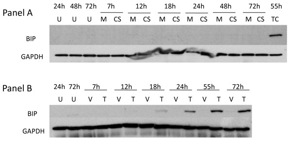
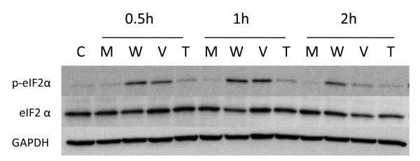
Results: We show that: 1) CS induces ER stress and activates components of the UPR; 2) reactive species in CS that promote oxidative stress are primarily responsible for UPR activation; 3) CS exposure results in increased expression of several genes with significant roles in attenuating oxidative stress; and 4) several major UPR regulators are increased either in expression (i.e., BiP and eIF2 alpha) or phosphorylation (i.e., phospho-eIF2 alpha) in a majority of human lung cancers.

Conclusion: These data indicate that chronic ER stress and recruitment of one or more UPR effector arms upon exposure to CS may play a pivotal role in the etiology or progression of lung cancers, and that phospho-eIF2 alpha and BiP may have diagnostic and/or therapeutic potential. Furthermore, we speculate that upregulation of UPR regulators (in particular BiP) may provide a pro-survival advantage by increasing resistance to cytotoxic stresses such as hypoxia and chemotherapeutic drugs, and that UPR induction is a potential mechanism that could be attenuated or reversed resulting in a more efficacious treatment strategy for lung cancer.


Figures

Figure 1
Figure 1
Induction of eIF2α phosphorylation in NHBE cells by cigarette smoke and thapsigargin. Panel A: NHBE cells were exposed to air (M = mock treatment) or 2R4F cigarette smoke with 35 cc puffs diluted in 250 cc air (CS = smoke treatment) for 20 minutes. Cells were then placed in fresh medium and returned to the incubator for the time periods specified. Western blots of whole cell lysates were probed with antibodies to phosphorylated eIF2α, eIF2α, and GAPDH as a loading control. C = untreated control. Panel B: NHBE cells were treated with either 1 uM thapsigargin in DMSO (T), or DMSO (VC = vehicle control) for the times specified. Western blots of whole cell lysates were probed with antibodies to phosphorylated eIF2α, eIF2α, and GAPDH. C = untreated control.
Figure 2
Figure 2
Dependence of eIF2α phosphorylation on PERK in A549 cells treated with cigarette smoke (and thapsigargin. Panel A: A549 cells were transfected with control siRNA (lanes 3 and 4) or PERK siRNA (lanes 5 and 6) as described in Methods. 24 h post-tranfection the cells were exposed to air (lanes 1, 3, and 5) or 2R4F cigarette smoke with 35 cc puffs diluted in 250 cc air (lanes 2,4, and 6) for 20 minutes. Cells were then placed in fresh medium and returned to the incubator for the time periods specified. Western blots of whole cell lysates were probed with antibodies to phosphorylated eIF2α, eIF2α, and GAPDH as a loading control. Panel B: A549 cells were transfected with control siRNA (lanes 3 and 4) or PERK siRNA (lanes 5 and 6) as described in Methods. 24 h post-tranfection the cells were treated with either 1 uM thapsigargin in DMSO (lanes 2,4, and 6) or DMSO (lanes 1, 3, and 5) for the times specified.
Figure 3
Figure 3
Effect of CS on ATF4 pathway. Panel A: NHBE cells were exposed to air (M = mock treatment) or 2R4F cigarette smoke with 35 cc puffs diluted in 250 cc air (CS = smoke treatment) for 20 minutes. Cells were then placed in fresh medium and returned to the incubator for the time periods specified. Nuclear extracts were prepared as described in the Methods section. Western blots of nuclear fractions were probed with antibodies to ATF4 and Lamin A/C, a nuclear antigen, as a loading control. Panel B: NHBE cells were exposed to air (M = mock treatment) or 2R4F cigarette smoke with 35 cc puffs diluted in 250 cc air (CS = smoke treatment) for 20 minutes. Cells were then placed in fresh medium and returned to the incubator for the time periods specified. Western blots of whole cell lysates were probed with antibodies to GADD34 and α-tubulin as a loading control. Panel C:A549 cells were exposed to air (M = mock treatment) or 2R4F cigarette smoke (CS = smoke treatment) for 20 minutes with 35 cc puffs diluted in 250 cc air. Cells were then placed in fresh medium and returned to the incubator for the time periods specified. Western blots of whole cell lysates were probed with antibodies to ATF3 and GAPDH.
Figure 4
Figure 4
Effect of CS on proteolytic cleavage of ATF6. Panel A: A549 cells were transfected with a plasmid expressing ATF6 (lanes 2 and 4, indicated by +), or a control plasmid (lanes 1 and 3), then treated with 2 mM DTT for 1 h (lanes 3 and 4) or left untreated (lanes 1 and 2). Western blots of whole cell lysates were probed with antibodies to ATF6 and α-tubulin. Panel B: All lanes show lysates from A549 cells transfected with a plasmid expressing ATF6. Cells were exposed to air (M = mock treatment) or 2R4F cigarette smoke (CS = smoke treatment) for 20 minutes with 35 cc puffs diluted in 250 cc air. Cells were then placed in fresh medium and returned to the incubator for the time periods specified. Western blots of whole cell lysates were probed with antibodies to ATF6 and α-tubulin. Gel bands were quantified as described in the Methods section. CS treatment resulted in a 23% and 26% decrease in ATF6 90 kDa protein at 2 and 4 h respectively (lane 5 compared to lane 6, lane 7 compared to lane 8).
Figure 5
Figure 5
Expression of BiP, XBP1, and grp94 in CS-treated human lung cells. NHBE cells were exposed to air (Mock), 2R4F cigarette smoke with 35 cc puffs diluted in 500 cc air (2R4F), or Brand B cigarette smoke with 35 cc puffs diluted in 500 cc air for 15 minutes. Cells were then placed in fresh medium and returned to the incubator for the time periods specified prior to RNA extraction. Four separate microarray representing four separate samples were used to analyze each condition. Mean intensity values are shown. * = p < 0.01 when compared to Mock.
Figure 6
Figure 6
Effect of thapsigargin and cigarette smoke on expression of BiP. Panel A: A549 cells were treated with 1 mM thapsigargin for the time periods specified. U = untreated cell control; V = vehicle (DMSO) control; T = thapsigargin. Panel B: A549 cells were cells were exposed to air (M = mock treatment) or 2R4F cigarette smoke (CS = smoke treatment) for 20 minutes (35 cc puffs were diluted in 250 cc air), after which the cells were placed in fresh medium and returned to the incubator for the time periods specified. Western blots of whole cell lysates were probed with antibodies to BiP and GAPDH. TC = thapsigargin treated cells as a positive internal control for Panel B.
Figure 7
Figure 7
BiP expression in normal human lung cells treated with CS long term treatment. NHBE cells were exposed to either 15% or 30% 2R4F cigarette smoke extract (CSE) prepared as described in (ref), or exposed to 2R4F cigarette smoke (CS) for 10 or 20 minutes (35 cc puffs were diluted in 250 cc air), after which the cells were immediately returned to the incubator without a media change for the time periods specified. C = untreated control.
Figure 8
Figure 8
Cigarette smoke inhibits XBP1 splicing. A549 cells were exposed to air (Panel A, mock treatment) or 2R4F cigarette smoke (Panel B) for 20 minutes with 35 cc puffs diluted in 250 cc air. Cells were then placed in fresh media with or without 1 uM thapsigargin or 10 μg/ml tunicamycin for the time periods specified, followed by cell lysis and RNA purification. Spliced and unspliced XBP1 mRNA was detected using PCR methodology. Spliced XBP1 is 398 base pairs and the unspliced variant is 434 base pairs. For each lane the extent of splicing was quantified as described in the Methods section and is presented in additional file 3 – Supplemental Table S3: Suppression of XBP1 splicing by cigarette smoke.
Figure 9
Figure 9
Relative effect of vapor and particulate ('tar') phases of cigarette smoke on eIF2α phosphorylation. NHBE cells were exposed to air (mock treatment M), whole cigarette smoke (W), vapor phase (V) or particulate 'tar' phase (T) from 2R4F cigarettes, after which the cells were placed in fresh medium and returned to the incubator for the time periods specified. Vapor phase was created by passing whole smoke through a Cambridge pad filter to trap the particulate matter. Particulate 'tar' phase was achieved by passing the whole smoke through 450 mg activated carbon to remove vapor phase components. Duration of whole smoke and vapor phase exposures was 20 minutes. Duration of particulate 'tar' phase exposures was 25 minutes in order to deliver an amount of particulate matter equivalent to that of the whole smoke exposure as some particulate matter is lost upon transit through the activated carbon. In all exposures the 35 cc puffs were diluted in 250 cc air. Western blots of whole cell lysates were probed with antibodies to phosphorylated eIF2α, eIF2α, and GAPDH.
Figure 10
Figure 10
Effect of antioxidant treatment on eIF2α phosphorylation following cigarette smoke exposure. A549 cells were exposed to air (mock treatment) or 2R4F cigarette smoke for 20 minutes with 35 cc puffs diluted in 250 cc air, with or without concurrent treatment with NAC or GSH (at 5 and 25 mM concentrations), after which the cells were placed in fresh medium and returned to the incubator for the time periods specified. Western blots of whole cell lysates were probed with antibodies to phosphorylated eIF2α and GAPDH.
Figure 11
Figure 11
Contribution of vapor and particulate phases of CS to XBP1 splicing inhibition in A549 cells. A549 cells were exposed to air (mock treatment), whole cigarette smoke (CS), vapor phase, or particulate 'tar' phase from 2R4F cigarettes as described in the legend for Figure 6, then placed in fresh media with or without 1 uM thapsigargin (THAP) and incubated for the time periods specified. PCR was used to determine the relative amounts of spliced and unspliced XBP1. For each lane the extent of splicing was quantified as described in the Methods section and is presented in additional file 3 – Supplemental Table S3: Suppression of XBP1 splicing by cigarette smoke.
Figure 12
Figure 12
Representative immunohistochemical expression features of phospho-eIF2α, eIF2α, and BiP proteins in different human lung lesions. The proteins assessed by immunohistochemistry are designated in rows, while the columns depict expression in normal lung tissues, representative positive (ISI values of > 6–9) and negative NSCLCs, and representative positive (ISI values of > 6–9) and negative Small Cell Carcinomas. Magnification, ×200.

References

    1. Lubin JH, Alavanja MC, Caporaso N, Brown LM, Brownson RC, Field RW, Garcia-Closas M, Hartge P, Hauptmann M, Hayes RB, Kleinerman R, Kogevinas M, Krewski D, Langholz B, Letourneau EG, Lynch CF, Malats N, Sandler DP, Schaffrath-Rosario A, Schoenberg JB, Silverman DT, Wang Z, Wichmann HE, Wilcox HB, Zielinski JM. Cigarette smoking and cancer risk: modeling total exposure and intensity. Am J Epidemiol. 2007;166:479–489. - PubMed
    1. Alberg AJ, Ford JG, Samet JM. Epidemiology of lung cancer: ACCP evidence-based clinical practice guidelines (2nd edition) Chest. 2007;132:29S–55S. - PubMed
    1. Albino AP, Huang X, Jorgensen E, Yang J, Gietl D, Traganos F, Darzynkiewicz Z. Induction of H2AX phosphorylation in pulmonary cells by tobacco smoke: a new assay for carcinogens. Cell Cycle. 2004;3:1062–1068. - PubMed
    1. Jorgensen ED, Dozmorov I, Frank MB, Centola M, Albino AP. Global Gene Expression Analysis of Human Bronchial Epithelial Cells Treated with Tobacco Condensates. Cell Cycle. 2004;3:1154–1168. - PubMed
    1. Albino AP, Huang X, Jorgensen ED, Gietl D, Traganos F, Darzynkiewicz Z. Induction of DNA double-strand breaks in A549 and normal human pulmonary epithelial cells by cigarette smoke is mediated by free radicals. Int J Oncol. 2006;28:1491–1505. - PubMed

Publication types

MeSH terms