. 2008 Aug 11:9:73.
doi: 10.1186/1471-2199-9-73.

Mutational analysis of highly conserved aspartate residues essential to the catalytic core of the piggyBac transposase

Affiliations

Mutational analysis of highly conserved aspartate residues essential to the catalytic core of the piggyBac transposase

James H Keith et al. BMC Mol Biol. .

Abstract

Background: The piggyBac mobile element is quickly gaining popularity as a tool for the transgenesis of many eukaryotic organisms. By studying the transposase which catalyzes the movement of piggyBac, we may be able to modify this vector system to make it a more effective transgenesis tool. In a previous publication, Sarkar A, Sim C, Hong YS, Hogan JR, Fraser MJ, Robertson HM, and Collins FH have proposed the presence of the widespread 'DDE/DDD' motif for piggyBac at amino acid positions D268, D346, and D447.

Results: This study utilizes directed mutagenesis and plasmid-based mobility assays to assess the importance of these residues as the catalytic core of the piggyBac transposase. We have functionally analyzed individual point-mutations with respect to charge and physical size in all three proposed residues of the 'DDD' motif as well as another nearby, highly conserved aspartate at D450. All of our mutations had a significant effect on excision frequency in S2 cell cultures. We have also aligned the piggyBac transposase to other close family members, both functional and non-functional, in an attempt to identify the most highly conserved regions and position a number of interesting features.

Conclusion: We found all the designated DDD aspartates reside in clusters of amino acids that conserved among piggyBac family transposase members. Our results indicate that all four aspartates are necessary, to one degree or another, for excision to occur in a cellular environment, but D450 seems to have a tolerance for a glutamate substitution. All mutants tested significantly decreased excision frequency in cell cultures when compared with the wild-type transposase.


Figures

Figure 1
Figure 1
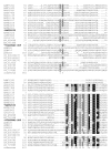
Alignment of piggyBac-related proteins. A BoxShade server alignment of the proteins listed in table 1. Residues aligning with piggyBac position 1–63 are shown. Only two piggyBac family proteins have been shown to catalyze transposition, these are indicated by bold face type and asterisks.
Figure 2
Figure 2
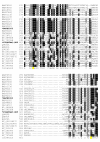
Alignment of piggyBac-related proteins. A BoxShade server alignment of the proteins listed in table 1. Residues aligning with piggyBac position 64–160 are shown. Only two piggyBac family proteins have been shown to catalyze transposition, these are indicated by bold face type and asterisks.
Figure 3
Figure 3
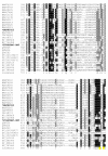
Alignment of piggyBac-related proteins. A BoxShade server alignment of the proteins listed in table 1. Residues aligning with piggyBac position 161–260 are shown. Only two piggyBac family proteins have been shown to catalyze transposition, these are indicated by bold face type and asterisks.
Figure 4
Figure 4
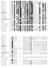
Alignment of piggyBac-related proteins. A BoxShade server alignment of the proteins listed in table 1. Residues aligning with piggyBac position 261–351 are shown. Only two piggyBac family proteins have been shown to catalyze transposition, these are indicated by bold face type and asterisks. The aspartate residues mutated in this study are highlighted in yellow.
Figure 5
Figure 5
Alignment of piggyBac-related proteins. A BoxShade server alignment of the proteins listed in table 1. Residues aligning with piggyBac position 352–451 are shown. Only two piggyBac family proteins have been shown to catalyze transposition, these are indicated by bold face type and asterisks. The aspartate residues mutated in this study are highlighted in yellow.
Figure 6
Figure 6
Alignment of piggyBac-related proteins. A BoxShade server alignment of the proteins listed in table 1. Residues aligning with piggyBac position 452–558 are shown. Only two piggyBac family proteins have been shown to catalyze transposition, these are indicated by bold face type and asterisks.
Figure 7
Figure 7
Alignment of piggyBac-related proteins. A BoxShade server alignment of the proteins listed in table 1. Residues aligning with piggyBac position 559 through the c-terminus are shown. Only two piggyBac family proteins have been shown to catalyze transposition, these are indicated by bold face type and asterisks.
Figure 8
Figure 8
Diagram of piggyBac ORF and genotypes used in this study. A diagrammatic representation of the piggyBac transposase mutant genotypes used in this study. The entire piggyBac ORF is displayed with the aspartates noted with their corresponding residue number. 4 × NLS represents 4 PSORT predicted nuclear localization patterns as described in discussion.
Figure 9
Figure 9
Schematic of the REN colony screen. (A) The donor plasmid pBKOα is co-transfected with one of the piggyBac expression plasmids (fig. 8), transformed into E. coli and plated. (B) Precise excision of the piggyBac cassette removes the lacZ open reading frame. Digestion with BglII removes any excess piggyBac expression plasmids and donor plasmids which have not undergone excision. Excision events are scored as white colonies. Plating a portion of undigested plasmids and scoring blue colonies yields the total number of potential donor plasmids.
Figure 10
Figure 10
Schematic of the blue/white colony excision assay. (A) The donor plasmid pCR2.1-piggyBac{SV40} is co-transfected with one of the piggyBac expression plasmids (fig. 8) transformed into E. coli and plated. (B) Precise excision of the piggyBac{SV40} cassette restores the original lacZ open reading frame at the original 'TTAA' insertion site. By plating on kanamycin media containing IPTG and X-gal, only E. coli containing donor plasmids will develop into colonies. Colonies which are blue represent excision events, while the number of blue and white colonies represents the total number of potential donor plasmids.
Figure 11
Figure 11
PCR analysis of donor plasmid sizes from the blue/white colony screen. PCR of 10 white (negative, top row) colonies and 10 blue (positive, bottom row) colonies, randomly chosen, from the blue/white colony screen is shown on a .9% TAE agarose gel stained with ethidium bromide. The amplicon crosses the entire donor fragment. Expected band size for the pre-excision plasmid is 1027 bp and 202 bp for the post-excision donor plasmid amplicon.
Figure 12
Figure 12
Relative frequency of excision obtained with the REN colony screen. Positive control, wild-type piggyBac, is set to 100. Data are expressed as a mean of three replicates +/- standard error bars. ANOVA test was performed using GraphPad Prism 3.0 software. Means were considered statistically significant when p-values less than 0.05 were obtained with the Dunnett's post-test. Statistical significance of difference with regards to positive control is indicated on all data figures as asterisks above bars. (p < 0.05)
Figure 13
Figure 13
Relative frequency of excision obtained with the Blue/White colony screen. Positive control, wild-type piggyBac, is set to 100. Data are expressed as a mean of three replicates +/- standard error bars. ANOVA test was performed using GraphPad Prism 3.0 software. Means were considered statistically significant when p-values less than 0.05 were obtained with the Dunnett's post-test. Statistical significance of difference with regards to positive control is indicated on all data figures as asterisks above bars. (p < 0.05)
Figure 14
Figure 14
Western blot of piggyBac mutant transposases. A western blot was performed on each piggyBac mutant with 125 μg total cell lysate per lane. The top row was probed with anti-piggyBac antibody and indicates the presence of the piggyBac transposase at 68 kDa in each mutant, the positive control, phsp-pBac, and a lack of transposase in the negative control. The bottom row was probed for actin, a 43 kDa protein, using anti-actin I-19 and shows equal loading in all lanes.

Similar articles

Cited by

References

    1. Rubin GM, Spradling AC. Genetic transformation of Drosophila with transposable element vectors. Science. 1982;218:348–353. doi: 10.1126/science.6289436. - DOI - PubMed
    1. Engels WR. P Elements in Drosophila. In: Saedler H, Gierl A, editor. Transposable Elements. Berlin , Springer; 1996. pp. 103–123.
    1. Craig NL. Mobile DNA II. Washington, D.C. , ASM Press; 2002. p. xviii, 1204 p., [32] p. of plates.
    1. Cary LC, Goebel M, Corsaro BG, Wang HG, Rosen E, Fraser MJ. Transposon mutagenesis of baculoviruses: analysis of Trichoplusia ni transposon IFP2 insertions within the FP-locus of nuclear polyhedrosis viruses. Virology. 1989;172:156–169. doi: 10.1016/0042-6822(89)90117-7. - DOI - PubMed
    1. Elick TA, Bauser CA, Fraser MJ. Excision of the piggyBac transposable element in vitro is a precise event that is enhanced by the expression of its encoded transposase. Genetica. 1996;98:33–41. doi: 10.1007/BF00120216. - DOI - PubMed

Publication types