. 2008 Aug;15(2):187-97.
doi: 10.1016/j.devcel.2008.07.004.

CP110 suppresses primary cilia formation through its interaction with CEP290, a protein deficient in human ciliary disease

Affiliations

CP110 suppresses primary cilia formation through its interaction with CEP290, a protein deficient in human ciliary disease

William Y Tsang et al. Dev Cell. 2008 Aug.

Abstract

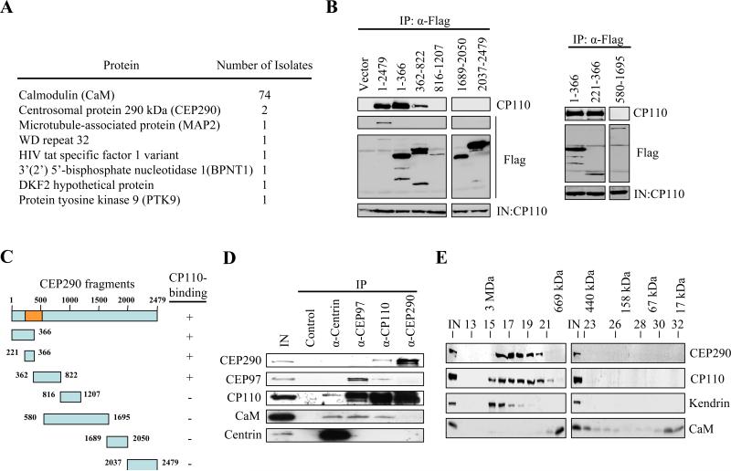
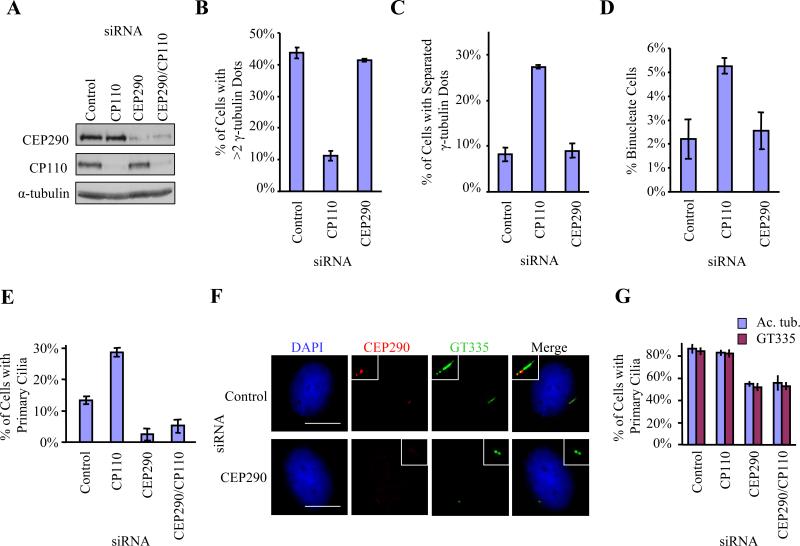
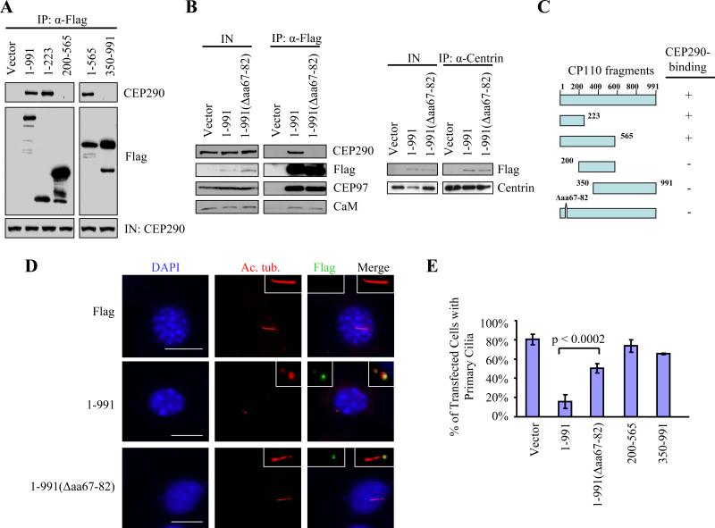
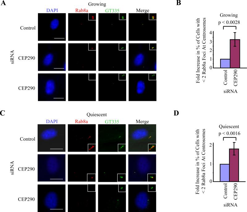
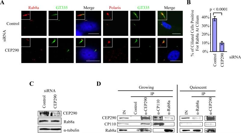
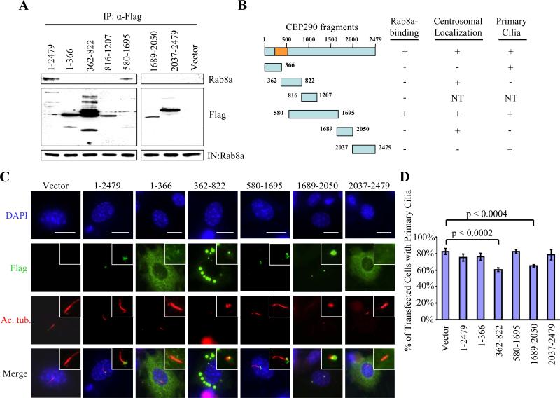
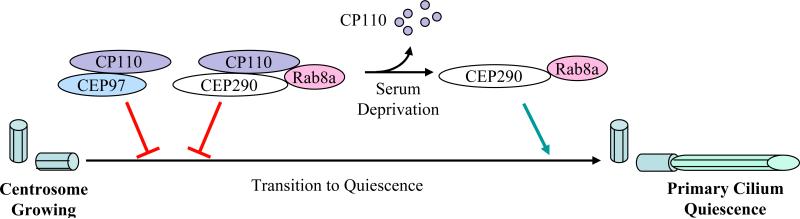
Primary cilia are nonmotile organelles implicated in signaling and sensory functions. Understanding how primary cilia assemble could shed light on the many human diseases caused by mutations in ciliary proteins. The centrosomal protein CP110 is known to suppress ciliogenesis through an unknown mechanism. Here, we report that CP110 interacts with CEP290--a protein whose deficiency is implicated in human ciliary disease--in a discrete complex separable from other CP110 complexes involved in regulating the centrosome cycle. Ablation of CEP290 prevents ciliogenesis without affecting centrosome function or cell-cycle progression. Interaction with CEP290 is absolutely required for the ability of CP110 to suppress primary cilia formation. Furthermore, CEP290 and CP110 interact with Rab8a, a small GTPase required for cilia assembly. Depletion of CEP290 interferes with localization of Rab8a to centrosomes and cilia. Our results suggest that CEP290 cooperates with Rab8a to promote ciliogenesis and that this function is antagonized by CP110.


Figures

Figure 1
Figure 1
CP110 interacts with CEP290 in vivo. (A) The number of times that each annotated protein was identified in the yeast two-hybrid screen is shown. (B) The indicated fragments of Flag-tagged CEP290 were expressed in 293T cells and immunoprecipitated from lysates. Flag-CEP290 fusion proteins and CP110 were detected after western blotting the resulting immunoprecipitates. Input CP110 was detected in lysates from each transfection (IN). (C) Summary of CEP290 truncation mutants and the results of in vivo binding experiments. The orange box denotes the CP110-binding domain based on the yeast two-hybrid screen. (D) Western blotting of endogenous CEP290, CEP97, CP110, CaM, and centrin after immunoprecipitation with anti-Flag (control), anti-centrin, anti-CEP97, anti-CP110, or anti-CEP290 antibodies from 293T cell extracts. IN represents input. (E) Cell extract was chromatographed on a Superose 6 gel filtration column, and the resulting fractions were blotted with antibodies against CP110, CEP290, kendrin, or CaM. Estimated molecular weights are indicated at the top of the panel.
Figure 2
Figure 2
RNAi-mediated suppression of CEP290 inhibits primary cilia formation. (A) Western blotting of CP110 and CEP290 in RPE-1 cells treated with control, CP110, CEP290, or both siRNAs. α-tubulin was used as a loading control. (B) U2OS cells were arrested at S phase with hydroxyurea for 48 hr after siRNA transfection. The percentages of cells with more than two γ-tubulin dots, indicative of centrosome over-duplication, were determined. (C) The percentages of U2OS cells with well-separated γ-tubulin dots, indicative of premature centrosome separation, were determined. (D) The percentages of binucleate WI38 cells, indicative of cytokinesis failure, were determined. (E) The percentages of growing RPE-1 cells with primary cilia were determined using acetylated tubulin as a marker. (F) RPE-1 cells transiently transfected with control or CEP290 siRNAs, induced to quiescence, stained with antibodies to glutamylated tubulin (GT335, green), CEP290 (red), and with DAPI (blue). Bar: 10 μM; insets: 2 μM. (G) The percentages of quiescent RPE-1 cells with primary cilia were determined using either acetylated tubulin (Ac. tub.) or gltuamylated tubulin (GT335) as a marker. In (B), (C), (D), (E) or (G), average data obtained from three independent experiments were shown. Error bars represent +/- S.D. About 200 cells for each siRNA condition were scored each time.
Figure 3
Figure 3
A CP110 mutant unable to bind CEP290 fails to inhibit primary cilia formation. (A) The indicated fragments of Flag-tagged CP110 were expressed in 293T cells and immunoprecipitated from lysates. Flag-CP110 fusion proteins and CEP290 were detected after western blotting the resulting immunoprecipitates. Input CEP290 was detected in lysates from each transfection (IN). (B) Left panel: Flag (vector), Flag-tagged full-length CP110 (1-991), and Flag-tagged CEP290-binding mutant of CP110 (1-991(Δaa67-82)) were expressed in 293T cells and immunoprecipitated from lysates. Flag-CP110 fusion proteins, CEP290, CEP97, and CaM were detected after blotting the resulting immunoprecipitates. IN represents input. Right panel: Western blot of Flag-CEP290 and centrin after immunoprecipitation with anti-centrin antibodies using extracts from 293T cells transfected with the indicated constructs. IN represents input. (C) Summary of CP110 truncation mutants and the results of in vivo binding experiments. (D) 3T3 cells transiently transfected with plasmids expressing the indicated constructs, induced to quiescence, and stained with antibodies to Flag (green), Ac. tub. (red), and with DAPI (blue). Bar: 10 μM; insets: 2 μM. (E) The percentages of transfected, quiescent 3T3 cells expressing primary cilia were determined using Ac. tub. as a marker. About 100 transfected cells were scored for each construct, and average data obtained from three independent experiments was shown. Error bars represent +/- S.D.
Figure 4
Figure 4
CEP290 recruits Rab8a to centrosomes. RPE-1 cells transfected with control or CEP290 siRNAs were (A, B) grown asynchronously in the presence of serum or (C, D) induced to quiescence, and stained with antibodies to gltuamylated tubulin (GT335, green), Rab8a (red), and with DAPI (blue). Bar: 10 μM; insets: 2 μM. (B, D) Histograms quantifying the fold increase in the percentage of cells with fewer than two Rab8a foci at centrosomes. About 100 cells were scored for each siRNA condition, and the histograms showed the averages of three independent experiments. Error bars represent +/- S.D. The average percentage of cells with fewer than two Rab8a foci after transfection with control siRNA was 16.5% in quiescent cells and 9.8% in growing cells.
Figure 5
Figure 5
CEP290 regulates entry of Rab8a into the primary cilium and interacts with Rab8a in vivo. (A) RPE-1 cells transfected with control or CEP290 siRNAs, induced to quiescence, and stained with antibodies to glutamylated tubulin (GT335, green), Rab8a or polaris (red), and with DAPI (blue). A ciliated cell is shown in each case. Bar: 10 μM; insets: 2 μM. (B) A histogram quantifying the percentage of ciliated cells that were positive for Rab8a at the cilium. About 100 cells from the ciliated population of cells treated with control or CEP290 siRNA were scored, and the averages of four independent experiments were shown. Error bars represent +/- S.D. (C) Western blotting of CEP290 and Rab8a in RPE-1 cells treated with control or CEP290 siRNAs. α-tubulin was used as a loading control. (D) Western blotting of endogenous CEP290, CP110 and Rab8a after immunoprecipitation with anti-Flag (control), anti-CEP290, anti-CP110, or anti-Rab8a antibodies from growing or quiescent 3T3 cell extracts. IN, input.
Figure 6
Figure 6
Ecotpic expression of certain CEP290 fragments prevents primary cilia formation. (A) The indicated fragments of Flag-tagged CEP290 were expressed in 293T cells and immunoprecipitated from lysates. Flag-CEP290 fusion proteins and Rab8a were detected after western blotting the resulting immunoprecipitates. Input Rab8a was detected in lysates from each transfection (IN). (B) Summary of CEP290 truncation mutants and the results of in vivo binding experiments. The orange box denotes the CP110-binding domain based on the yeast two-hybrid screen. NT denotes “not tested.” (C) 3T3 cells transiently transfected with plasmids expressing Flag-CEP290 truncation mutants, induced to quiescence, and stained with antibodies to Flag (green), Ac. tub. (red), and with DAPI (blue). Bar: 10 μM; insets: 2 μM. (D) The percentages of transfected, quiescent 3T3 cells expressing primary cilia were determined using Ac. tub. as a marker. About 100 transfected cells for each construct were scored, and average data obtained from three independent experiments were shown. Error bars represent +/- S.D.
Figure 7
Figure 7
A model depicting the role of CEP290 in mediating ciliogenesis. In growing cells, CP110 antagonizes the action of CEP290, which in turn prevents CEP290-dependent Rab8a ciliogenesis. Another complex containing CP110 and CEP97 also serves to suppress primary cilia assembly. When cells exit the cell cycle, the levels of CP110 diminish dramatically. This frees up CEP290, which in turn cooperates with Rab8a to promote primary cilia formation.

References

    1. Andersen JS, Wilkinson CJ, Mayor T, Mortensen P, Nigg EA, Mann M. Proteomic characterization of the human centrosome by protein correlation profiling. Nature. 2003;426:570–574. - PubMed
    1. Baala L, Audollent S, Martinovic J, Ozilou C, Babron MC, Sivanandamoorthy S, Saunier S, Salomon R, Gonzales M, Rattenberry E, et al. Pleiotropic effects of CEP290 (NPHP6) mutations extend to Meckel syndrome. Am. J. Hum. Genet. 2007;81:170–179. - PMC - PubMed
    1. Badano JL, Teslovich TM, Katsanis N. The centrosome in human genetic disease. Nat. Rev. Genet. 2005;6:194–205. - PubMed
    1. Bettencourt-Dias M, Glover DM. Centrosome biogenesis and function: centrosomics brings new understanding. Nat. Rev. Mol. Cell Biol. 2007;8:451–463. - PubMed
    1. Chang B, Khanna H, Hawes N, Jimeno D, He S, Lillo C, Parapuram SK, Cheng H, Scott A, Hurd RE, et al. In-frame deletion in a novel centrosomal/ciliary protein CEP290/NPHP6 perturbs its interaction with RPGR and results in early-onset retinal degeneration in the rd16 mouse. Hum. Mol. Genet. 2006;15:1847–1857. - PMC - PubMed

Publication types

MeSH terms