. 2008 Aug;15(2):248-60.
doi: 10.1016/j.devcel.2008.06.013.

Crossveinless-2 Is a BMP feedback inhibitor that binds Chordin/BMP to regulate Xenopus embryonic patterning

Affiliations

Crossveinless-2 Is a BMP feedback inhibitor that binds Chordin/BMP to regulate Xenopus embryonic patterning

Andrea L Ambrosio et al. Dev Cell. 2008 Aug.

Abstract

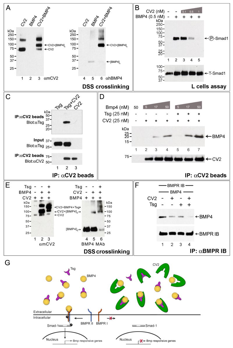
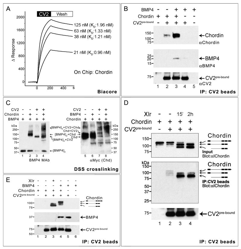
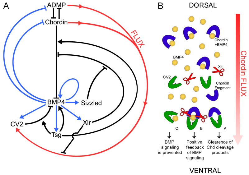
Vertebrate Crossveinless-2 (CV2) is a secreted protein that can potentiate or antagonize BMP signaling. Through embryological and biochemical experiments we find that: (1) CV2 functions as a BMP4 feedback inhibitor in ventral regions of the Xenopus embryo; (2) CV2 complexes with Twisted gastrulation and BMP4; (3) CV2 is not a substrate for tolloid proteinases; (4) CV2 binds to purified Chordin protein with high affinity (K(D) in the 1 nM range); (5) CV2 binds even more strongly to Chordin proteolytic fragments resulting from Tolloid digestion or to full-length Chordin/BMP complexes; (6) CV2 depletion causes the Xenopus embryo to become hypersensitive to the anti-BMP effects of Chordin overexpression or tolloid inhibition. We propose that the CV2/Chordin interaction may help coordinate BMP diffusion to the ventral side of the embryo, ensuring that BMPs liberated from Chordin inhibition by tolloid proteolysis cause peak signaling levels.


Figures

Figure 1
Figure 1. CV2 is a Secreted BMP Feedback Inhibitor
(A) Normal CV2 expression at stage 22 (hemisection). vb, ventral blastopore (B) CV2MO microinjection increases CV2 expression. (C) The CV2 negative feedback loop requires BMP4. For each experimental sample at least 25 embryos were examined, with similar results. (D) qRT-PCR showing increased expression of the ventral marker Vent1 in CV2-depleted embryos. (E) CV2 depletion reduces expression of the dorsal/forebrain marker Six3. (F) Endogenous Smad1 phosphorylation is increased by CV2 depletion at gastrula and neurula stages 12, 13 and 14. A pSmad1 signal was detectable in the stage 14 uninjected lane upon longer exposure. Total Smad1 antibody (T-Smad1) staining was used as loading control.
Figure 2
Figure 2. CV2 and Chordin Compensate for Each Other in Xenopus D-V Patterning
(A) Uninjected embryo showing CV2 expression, which is used here as a BMP4 signaling readout (n=17). Inset shows mid-gastrula embryo stained for Chordin (Chd) and Sizzled (Szl) (n=24). (B) CV2 depletion upregulates its own expression (n=15), as well as increasing Szl and decreasing Chd (n=20). (C) Depletion of the BMP antagonist Chordin also increases the ventral CV2 and Szl expression domains (n=13 and n=18, respectively). (D) When co-injected, CV2 MO and Chd MO show a marked expansion of the CV2 and Szl expression domains (n=15 and n=25, respectively). (E–H) qRT-PCR analyses of single and double CV2 and Chd morphants for the D-V markers Szl, CV2, Gsc and Chd at late gastrula stage 12.5. (I) Endogenous Smad1 phosphorylation is increased by co-injection of Chd MO and CV2 MO in stage 11 embryos. (J) The CV2 cleavage sequence contains the conserved low-pH GDPH autocatalytic site present in mucins. hMuc2, human mucin-2. (K) Chordin, but not full-length CV2, is cleaved by the extracellular zinc-metalloproteinases Xolloid-related (Xlr) and BMP1 (lanes 1–6). However, cleavage of full-length CV2 (80 kD band) is triggered by low pH (lanes 9 and 10). (L–N) Ventral injections of mRNAs encoding full-length CV2 (CV2-FL, n=56, of which 95% had partial secondary axes, three independent experiments), N-terminal CV2 fragment terminating at the GDPH cleavage site (CV2 N-Ter, n=42, no secondary axes observed), or a secreted C-terminal fragment encoding most of the vWFd domain (CV2 C-Ter, n=45, no secondary axes observed). The insets show injected embryos at late neurula stage hybridized with the pan-neural marker Sox2.
Figure 3
Figure 3. Twisted-Gastrulation (Tsg) is Required for the Effects of CV2 Loss-of-Function and Overexpression
(A) Simultaneous depletion of CV2 and Chordin strikingly increased CV2 expression, reflecting increased BMP signaling (see Figure S5 for controls). (B) The effects of CV2 MO and Chd MO require Tsg activity (pro-BMP effect of Tsg). (C) CV2 protein injection into the blastula cavity induces strong dorsalization of the Xenopus embryo, as indicated by the expansion of the dorsal markers Xag1, Six3, and Krox20 (n=12, all strongly dorsalized). Inset shows an uninjected embryo. (D) CV2 protein requires endogenous Tsg for its anti-BMP activity (n=10, all embryos similarly affected). Inset shows Tsg MO injected embryo. (E) CV2 protein injection expands the neural tube (n=17). Inset shows uninjected embryo. (F) Tsg and CV2 protein co-injection renders CV2 a stronger BMP antagonist, expanding the nervous system marked by Sox3 (n=16, all co-injected embryos were more dorsalized than those injected with CV2 protein alone despite some individual variations). Inset shows embryo injected with Tsg protein alone.
Figure 4
Figure 4. CV2, BMP4 and Tsg form a Ternary Complex that Inhibits BMP Signaling
(A) DSS crosslinking showing that CV2 binds BMP4 directly. (B) CV2 dose-dependently blocks BMP4-induced phosphorylation of endogenous Smad1 in mouse L-cells; BMP4 was added for 30 min in serum-free medium after pre-incubation with CV2 protein for 2 hours at 4°C. (C) Co-immunoprecipitation demonstrating that purified CV2 binds Tsg. (D) Tsg facilitates the binding of CV2 to BMP4. (E) CV2, BMP4 and Tsg form a ternary complex. Left panel shows DSS crosslinking products stained with anti-CV2 antibody. Right panel, same samples examined with anti-BMP monoclonal antibody (anti-Tsg antibody showed smears due to crosslinking of Tsg with itself). Tsg protein alone is not recognized by either antibody (not shown). (F) CV2 and Tsg additively block the binding of BMP4 to BMPR-1b-Fc. (G) Model of the molecular interactions of CV2, Tsg and BMP4 in a ternary complex.
Figure 5
Figure 5. CV2 and Chordin Bind to Each Other
(A) Biacore sensograms of the CV2-Chordin interaction in real-time showing an average KD of 1.37 nM. The time at which the binding of CV2 protein stops and the buffer wash starts is indicated. (B) Co-immunoprecipitation in solution showing that Chordin and BMP4 bind better to CV2 in the presence of each other. Top panel shows Western blot immunostained with anti-Chordin, middle panel with monoclonal anti-BMP4, and bottom panel (loading control) with anti-CV2 antibody. (C) CV2, Chordin and BMP4 form a ternary complex. Left panel shows crosslinked products stained with BMP4 monoclonal antibody; the antigenicity of BMP4 in crosslinked complexes with Chd and CV2 increases (the unbound BMP4 dimer band indicates the amount of BMP4 protein present in the complexes). The right panel the same products stained for myc-tagged Xenopus Chordin protein. The position of the BMP4/CV2/Chordin ternary complex is indicated. (D) CV2 binds better to Chordin cleavage products than to Chordin full-length protein. Some cleavage products were present in the Chordin protein preparation, and these were enriched in lane 2. (E) BMP4 pre-bound to Chordin enhances the binding preference of CV2 beads for full-length Chordin (lane 4). At this exposure level the binding of full-length Chordin to CV2 is undetectable (lane 2), but is greatly increased by adding Xlr or BMP4 (lanes 3 and 4).
Figure 6
Figure 6. The pro-BMP Function of CV2 is Revealed in Epistatic Experiments with Chordin or Tolloid
(A) Expression of the eye field marker Rx2a in uninjected Xenopus late neurula embryo (n=45), anterior view. (B) Chordin protein injection (2 μM, 60 nl) into the blastocoele at late blastula (stage 9.5) caused dorsalization and an increase in Rx2a expression (n=54). (C) CV2-depleted hosts were more sensitive to the anti-BMP effects of Chordin, as indicated by the expansion in the Rx2a domain (n=48). (D) Uninjected early neurula (stage 13, side view) showing Otx2 expression in the future forebrain and midbrain regions (n=19). (E) Chordin protein injection expands Otx2 in wild-type embryos (n=25). (F) CV2 depletion sensitizes the embryo to the effects of Chordin on Otx2 (n=23). Note that the border of Otx2 expression expands posteriorly. (G–H) qRT-PCR analysis of the D-V markers Chd, CV2 and Szl after Chordin protein injection into wild-type and CV2-depleted embryos at late blastula. The bars indicate standard deviation between two groups of seven embryos each. (J and K) Anterior views of uninjected control or embryo microinjected four times with 250 pg of DN-Xlr mRNA, which inhibits the proteolytic degradation of Chordin. Note that the Otx2-positive forebrain (fb), midbrain, and cement gland (cg) regions are expanded, consistent with the anti-BMP effects of Tolloid inhibition (n=27). (L) In CV2 depleted embryos Otx2 expression is greatly expanded by DN-Xlr mRNA (n=27). The dotted line indicates the eye field (eye), which is more weakly stained by Otx2.
Figure 7
Figure 7. Model of the Molecular Interaction of CV2, Chordin, Tsg and BMP4
(A) Model of the regulation of D-V patterning by a network of extracellular proteins secreted by the dorsal and ventral centers of the Xenopus gastrula. Arrows in black indicate direct protein-protein interactions in the extracellular space, blue arrows transcriptional regulation by the BMP-responsive transcription factors Smad1/5/8, and the red arrow the hypothetical flux of Chordin/ADMP/BMP from the dorsal toward the ventral center of the embryo, where it would bind to CV2. This model of D-V patterning is self-regulating because at low BMP levels the transcription of the BMP-like molecule ADMP is activated, and at high-BMP levels the BMP antagonist CV2 and the tolloid inhibitor Sizzled are upregulated (Reversade and De Robertis, 2005; Lee et al., 2006). The function of Tsg is to both increase BMP inhibition by CV2 and Chd and to promote BMP4 signaling in their absence. The tolloid protease Xlr cleaves Chordin/ADMP/BMP complexes, releasing active BMPs concentrated on the ventral side. (B) Model in which Chordin flow would help transport BMPs and Chordin from the dorsal to the ventral side of the Xenopus embryo. Three possible outcomes are indicated.

Comment in

Similar articles

Cited by

References

    1. Ben-Zvi D, Shilo BZ, Fainsod A, Barkai N. Scaling of the BMP activation gradient in Xenopus embryos. Nature. 2008 in press. - PubMed
    1. Binnerts ME, Wen X, Cante-Barrett K, Bright J, Chen HT, Asundi V, Sattari P, Tang T, Boyle B, Funk W, Rupp F. Human Crossveinless-2 is a novel inhibitor of bone morphogenetic proteins. Biochem Biophys Res Commun. 2004;315:272–280. - PubMed
    1. Blair SS. Wing vein patterning in Drosophila and the analysis of intercellular signaling. Annu Rev Cell Dev Biol. 2007;23:293–319. - PubMed
    1. Blitz IL, Chow KW, Chang C. Twisted gastrulation loss-of-function analyses support its role as a BMP inhibitor during early Xenopus embryogenesis. Development. 2003;130:4975–4988. - PubMed
    1. Blitz IL, Shimmi O, Wünnenberg-Stapleton K, O’Connor MB, Cho KW. Is chordin a long-range- or short-range-acting factor? Roles for BMP1-related metalloproteases in chordin and BMP4 autofeedback loop regulation. Dev Biol. 2000;223:120–138. - PubMed

Publication types

MeSH terms