

Subacute neural stem cell therapy for traumatic brain injury
- PMID: 18694578
- PMCID: PMC2874889
- DOI: 10.1016/j.jss.2008.03.037
Subacute neural stem cell therapy for traumatic brain injury
Abstract
Introduction: Traumatic brain injury (TBI) frequently results in devastating and prolonged morbidity. Cellular therapy is a burgeoning field of experimental treatment that has shown promise in the management of many diseases, including TBI. Previous work suggests that certain stem and progenitor cell populations migrate to sites of inflammation and improve functional outcome in rodents after neural injury. Unfortunately, recent study has revealed potential limitations of acute and intravenous stem cell therapy. We studied subacute, direct intracerebral neural stem and progenitor cell (NSC) therapy for TBI.
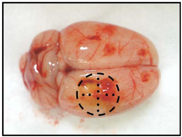
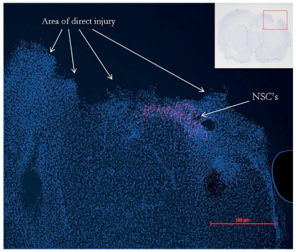
Materials and methods: The NSCs were characterized by flow cytometry and placed (400,000 cells in 50 muL 1x phosphate-buffered saline) into and around the direct injury area, using stereotactic guidance, of female Sprague Dawley rats 1 wk after undergoing a controlled cortical impact injury. Immunohistochemistry was used to identify cells located in the brain at 48 h and 2 wk after administration. Motor function was assessed using the neurological severity score, foot fault, rotarod, and beam balance. Cognitive function was assessed using the Morris water maze learning paradigm. Repeated measures analysis of variance with post-hoc analysis were used to determine significance at P < 0.05.
Results: Immunohistochemistry analysis revealed that 1.4-1.9% of infused cells remained in the neural tissue at 48 h and 2 wk post placement. Nearly all cells were located along injection tracks at 48 h. At 2 wk some cell dispersion was apparent. Rotarod motor testing revealed significant increases in maximal speed among NSC-treated rats compared with saline controls at d 4 (36.4 versus 27.1 rpm, P < 0.05) and 5 (35.8 versus 28.9 rpm, P < 0.05). All other motor and cognitive evaluations were not significantly different compared to controls.
Conclusions: Placement of NSCs led to the cells incorporating and remaining in the tissues 2 wk after placement. Motor function tests revealed improvements in the ability to run on a rotating rod; however, other motor and cognitive functions were not significantly improved by NSC therapy. Further examination of a dose response and optimization of placement strategy may improve long-term cell survival and maximize functional recovery.
Figures


References
-
- Kraus JF, Fife D, Conroy C. Pediatric brain injuries: The nature, clinical course, and early outcomes in a defined United States' population. Pediatrics. 1987;79:501. - PubMed
-
- Keel M, Trentz O. Pathophysiology of polytrauma. Injury. 2005;36:691. - PubMed
-
- Consensus conference. Rehabilitation of persons with traumatic brain injury. NIH Consensus Development Panel on Rehabilitation of Persons with Traumatic Brain Injury. JAMA. 1999;282:974. - PubMed
-
- Doppenberg EM, Choi SC, Bullock R. Clinical trials in traumatic brain injury: Lessons for the future. J Neurosurg Anesthesiol. 2004;16:87. - PubMed
-
- Sinson G, Voddi M, McIntosh TK. Combined fetal neural transplantation and nerve growth factor infusion: Effects on neurological outcome following fluid-percussion brain injury in the rat. J Neurosurg. 1996;84:655. - PubMed
Publication types
MeSH terms
Grants and funding
LinkOut - more resources
Full Text Sources
Other Literature Sources
