. 2008 Nov 28;383(5):1081-96.
doi: 10.1016/j.jmb.2008.07.071. Epub 2008 Jul 30.

Variable oligomerization modes in coronavirus non-structural protein 9

Affiliations

Variable oligomerization modes in coronavirus non-structural protein 9

Rajesh Ponnusamy et al. J Mol Biol. .

Abstract

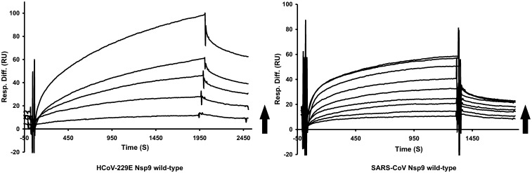
Non-structural protein 9 (Nsp9) of coronaviruses is believed to bind single-stranded RNA in the viral replication complex. The crystal structure of Nsp9 of human coronavirus (HCoV) 229E reveals a novel disulfide-linked homodimer, which is very different from the previously reported Nsp9 dimer of SARS coronavirus. In contrast, the structure of the Cys69Ala mutant of HCoV-229E Nsp9 shows the same dimer organization as the SARS-CoV protein. In the crystal, the wild-type HCoV-229E protein forms a trimer of dimers, whereas the mutant and SARS-CoV Nsp9 are organized in rod-like polymers. Chemical cross-linking suggests similar modes of aggregation in solution. In zone-interference gel electrophoresis assays and surface plasmon resonance experiments, the HCoV-229E wild-type protein is found to bind oligonucleotides with relatively high affinity, whereas binding by the Cys69Ala and Cys69Ser mutants is observed only for the longest oligonucleotides. The corresponding mutations in SARS-CoV Nsp9 do not hamper nucleic acid binding. From the crystal structures, a model for single-stranded RNA binding by Nsp9 is deduced. We propose that both forms of the Nsp9 dimer are biologically relevant; the occurrence of the disulfide-bonded form may be correlated with oxidative stress induced in the host cell by the viral infection.


Figures

Fig. 1
Fig. 1
Superimposition of monomers. Ribbon representation of HCoV-229E Nsp9 wild-type (green) and Cys69Ala mutant (red) monomers, superimposed with a Cα r.m.s. deviation of 0.71 Å. Loop L23 of wild-type HCoV-229E Nsp9 could not be built due to the lack of electron density.
Fig. 2
Fig. 2
Structural features of the homodimers of wild-type HCoV-229E Nsp9 and the Cys69Ala mutant; the two monomers are colored red and green, respectively; the disulfide, where present, is shown in yellow. N and C denote the amino and carboxy termini, respectively, of the polypeptide chains. (a) Ribbon representation of the disulfide-linked wild-type HCoV-229E Nsp9 dimer. (b) Residues involved in the dimer interface of wild-type Nsp9 (sticks; red, oxygen; blue, nitrogen; yellow, carbon). Intermolecular hydrogen bonds are indicated by broken lines. (c) HCoV-229E Cys69Ala mutant dimer. (d) Residues involved in the dimer interface of the mutant Nsp9. Color code is the same as in b. The closest approach between the C-terminal α-helices, between the Cα atoms of Gly A100 and Ala B97, is indicated by a dotted line.
Fig. 3
Fig. 3
Ribbon representation of the wild-type HCoV-229E Nsp9 hexamer. Three dimers of the protein form a hexamer through the 32 axis of symmetry. The threefold axis is at the center of the hexamer. Nsp9 monomers in the upper layer are colored red, blue and green, and those in the lower layer are colored yellow, cyan and magenta. The two sulfate ions on the threefold axis are indicated in the same colors. Each sulfate is three-fold disordered. The twofold axes run between the monomers.
Fig. 4
Fig. 4
Nsp9 crosslinking using glutaraldehyde. Crosslinking was carried out with different concentrations of protein (10–100 μM) using 0.01% (v/v) glutaraldehyde. The molecular mass of the cross-linking products is indicated. Wild-type SARS-CoV Nsp9 and the HCoV-229E Nsp9 Cys69Ala mutant form higher oligomers at a protein concentration of 100 μM, presumably involving interactions similar to those seen in the crystal structure. In contrast, wild-type HCoV-229E Nsp9 does not form oligomers higher than trimers.
Fig. 5
Fig. 5
Gel mobility-shift assay (zone-interference gel electrophoresis, 1% agarose; see Materials and Methods) probing oligonucleotide binding to Nsp9. (a) Wild-type HCoV-229E Nsp9; (b) wild-type SARS-CoV Nsp9. Lane 1, protein without ssDNA; lane 2, 24-mer dsDNA; lanes 3–10, various lengths of ssDNA from 6-mer to 50-mer. Wild-type HCoV-229E Nsp9 displays a linear increase of the shift with increasing length of ssDNA, whereas the increase is step-wise for SARS-CoV Nsp9. (c) Gel mobility-shift analysis for mutant proteins, compared to the corresponding wild-type. Lanes 1, protein without ssDNA; lanes 2, 24-mer, and lanes 3, 55-mer ssDNA with protein. The HCoV-229E Nsp9 Cys69Ala mutant (HAM) and the Cys69Ser mutant (HSM) do not show any shift with the 24-mer (lane 2) and only a small shift with the 55-mer (lane 3), whereas the SARS-CoV Nsp9 Cys73Ala mutant (SAM) and the Cys73Ser mutant (SSM) exhibit shifts with the 55-mer oligonucleotide that are similar to wild-type SARS-CoV Nsp9. The upper bands (gray) in lanes 3 for HAM and HSM correspond to precipitated, unbound 55-mer oligonucleotide (not stained by Coomassie brilliant blue; see Materials and Methods).
Fig. 6
Fig. 6
HCoV-229E Nsp9 binding to ssDNA analyzed by surface plasmon resonance, in the presence and in the absence of DTT. A 5′-biotinylated 50-mer oligonucleotide was immobilized to an SA chip up to 88 RU. a and b, The binding curves for a 20 μM fresh preparation of the Nsp9 Cys69Ala mutant and for wild-type Nsp9, respectively, both injected in the presence of 5 mM DTT. c, The binding curve for a 20 μM aged preparation of wild-type Nsp9, injected in the absence of 5 mM DTT.
Fig. 7
Fig. 7
Oligomers of SARS-CoV Nsp9 (PDB codes 1QZ8 (a) and 1UW7 (b)) and the HCoV-229E Nsp9 Cys69Ala mutant (c). Independent of space group symmetry (1QZ8, P6122; 1UW7, P4322; and HCoV-229E Nsp9 Cys69Ala mutant, P212121), two common dimer interfaces are present in these crystal structures of Nsp9. One interface is formed mainly by the C-terminal α-helix (red), and the other by strand β5 (blue). We propose that the ssRNA (black) could wrap around the Nsp9 polymer by forming a left-handed helix (a), with approximately 40 nucleotides bound per Nsp9 dimer.
Supplementary data
Supplementary data
Supplementary data
Supplementary data
Supplementary data
Supplementary data
Supplementary data
Supplementary data
Supplementary data
Supplementary data
Supplementary data
Supplementary data
Supplementary data
Supplementary data

References

    1. Drosten C., Gunther S., Preiser W., van der Werf S., Brodt H.R., Becker S. Identification of a novel coronavirus in patients with severe acute respiratory syndrome. N. Engl. J. Med. 2003;348:1967–1976. - PubMed
    1. Ksiazek T.G., Erdman D., Goldsmith C.S., Zaki S.R., Peret T., Emery S. A novel coronavirus associated with severe acute respiratory syndrome. N. Engl. J. Med. 2003;348:1953–1966. - PubMed
    1. Kuiken T., Fouchier R.A., Schutten M., Rimmelzwaan G.F., van Amerongen G., van Riel D. Newly discovered coronavirus as the primary cause of severe acute respiratory syndrome. Lancet. 2003;362:263–270. - PMC - PubMed
    1. Peiris J.S., Chu C.M., Cheng V.C., Chan K.S., Hung I.F., Poon L.L. Clinical progression and viral load in a community outbreak of coronavirus-associated SARS pneumonia: a prospective study. Lancet. 2003;361:1767–1772. - PMC - PubMed
    1. Groneberg D.A., Hilgenfeld R., Zabel P. Molecular mechanisms of severe acute respiratory syndrome (SARS) Respir. Res. 2005;6:8–23. - PMC - PubMed

Publication types

MeSH terms