. 2008 Oct;28(20):6262-77.
doi: 10.1128/MCB.00923-08. Epub 2008 Aug 11.

Protein arginine methyltransferase 5 suppresses the transcription of the RB family of tumor suppressors in leukemia and lymphoma cells

Affiliations

Protein arginine methyltransferase 5 suppresses the transcription of the RB family of tumor suppressors in leukemia and lymphoma cells

Li Wang et al. Mol Cell Biol. 2008 Oct.

Abstract

The proper epigenetic modification of chromatin by protein arginine methyltransferases (PRMTs) is crucial for normal cell growth and health. The human SWI/SNF-associated PRMT5 is involved in the transcriptional repression of target genes by directly methylating H3R8 and H4R3. To further understand the impact of PRMT5-mediated histone methylation on cancer, we analyzed its expression in normal and transformed human B lymphocytes. Our findings reveal that PRMT5 protein levels are enhanced in various human lymphoid cancer cells, including transformed chronic lymphocytic leukemia (B-CLL) cell lines. PRMT5 overexpression is caused by the altered expression of the PRMT5-specific microRNAs 19a, 25, 32, 92, 92b, and 96 and results in the increased global symmetric methylation of H3R8 and H4R3. An evaluation of both epigenetic marks at PRMT5 target genes such as RB1 (p105), RBL1 (p107), and RBL2 (p130) showed that promoters H3R8 and H4R3 are hypermethylated, which in turn triggers pocket protein transcriptional repression. Furthermore, reducing PRMT5 expression in WaC3CD5 B-CLL cells abolishes H3R8 and H4R3 hypermethylation, restores RBL2 expression, and inhibits cancer cell proliferation. These results indicate that PRMT5 overexpression epigenetically alters the transcription of key tumor suppressor genes and suggest a causal role of the elevated symmetric methylation of H3R8 and H4R3 at the RBL2 promoter in transformed B-lymphocyte pathology.


Figures
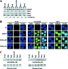
FIG. 1.
FIG. 1.
PRMT5 is overexpressed in transformed B-cell lines. (A) Western blot analysis of PRMT5 and human SWI/SNF subunits using 20 μg of nuclear (N) and cytosolic (C) extracts from either normal cells or the indicated transformed cell lines. The expression of α-tubulin was measured to discern between N and C fractions. (B) Immunofluorescence was used to measure the levels of PRMT5, H3(Me2)R8, and H4(Me2)R3 in normal and transformed B-CLL cells. Fluorescein isothiocyanate-labeled goat anti-rabbit antibody was used to detect PRMT5 and modified histones, and DAPI was used to stain nuclei. Pictures were taken at 100× magnification. PI, preimmune antibody. (C) Western blot analysis was carried using 2 μg of total histones isolated from either normal or transformed B cells when the symmetric methylation of H3R8 was evaluated. When anti-H4(Me2)R3 was used to monitor the methylation of H4R3, 7.5 μg of total histones was loaded on the gel. Ponceau S staining is included to show that all four core histones were transferred equally to the nitrocellulose membrane.
FIG. 2.
FIG. 2.
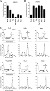
Expression of PRMT5 is enhanced at the translational level in transformed lymphoid cells. (A) To measure the steady-state levels of PRMT5 mRNA in normal and transformed B cells, real-time RT-PCR was performed three times in triplicates using total RNA. GAPDH mRNA levels were used as internal controls. (B) To verify that other members of the PRMT family also are transcriptionally repressed, we measured the levels of PRMT6 by real-time RT-PCR as described for panel A. (C) Representative polyribosome profiles from normal and transformed B-CLL cells were generated by loading and fractionating whole-cell lysates on 15 to 40% sucrose gradients. The optical density of each fraction was determined at 254 nm (OD254), and the position of 40S, 60S, and 80S mRNA and polyribosomes is indicated (upper panel). The lower panels show the level of PRMT5 mRNA present in each fraction, as measured by real-time RT-PCR using total RNA precipitated from each gradient fraction. β-actin mRNA was used as an internal control to normalize PRMT5 mRNA levels in each fraction. The data points in each graph represent the averages from triplicate RT-PCRs ± standard deviations.
FIG. 3.
FIG. 3.
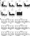
PRMT5-specific miRNAs are expressed differently in normal and transformed B cells, and their reexpression can inhibit PRMT5 translation. (A) RPAs were performed on 10 μg of total RNA isolated from the indicated cells using miR-19a, miR-19b, miR-25, miR-32, miR-92, miR-92b, and miR-96 probes. Mature and protected miRNA bands were quantitated from three independent experiments using ImageQuant v5.0 and were plotted as bar graphs. (B) WaC3CD5 and Mec1 cell lines were electroporated with either 2.5 or 5.0 μg of the indicated wild-type (WT) and mutant (MUT) dsRNAs, and 20 μg of whole-cell extracts was analyzed by Western blotting using anti-PRMT5 antibody. To show that samples were equally loaded, β-actin levels were measured in each sample. To demonstrate the specificity of each miRNA, miR-197, which does not exhibit any sequence complementarity to the PRMT5 3′UTR, is shown.
FIG. 4.
FIG. 4.
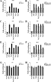
PRMT5 is recruited to and epigenetically modifies the RB family of tumor suppressor genes. ChIP assays were performed on cross-linked chromatin from either normal or transformed B cells using preimmune (PI) or immune anti-PRMT5 (A, C, and E) antibody or using immune anti-H3(Me2)R8 or anti-H4(Me2)R3 antibody (B, D, and F). Immunopurified DNA was amplified by real-time PCR using RB1-, RBL1-, or RBL2-specific primer sets and probes. (G and H) To show that PRMT5 recruitment to the RB family of tumor suppressors is specific, we examined the PRMT5 association with the promoter of ST5. The change in enrichment with each antibody was calculated relative to that of the PI sample, and each ChIP experiment was repeated twice in triplicate.
FIG. 5.
FIG. 5.
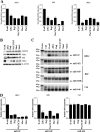
Expression of pocket proteins in normal and transformed B cells. (A) The mRNA expression of the RB family of tumor suppressors was measured in normal and transformed B cells by real-time RT-PCR. Graphs show normalized changes in expression for each gene relative to that of normal B cells using GAPDH as an internal control. (B) Whole-cell extracts were prepared from normal and transformed B cells, and 25 μg of total protein was analyzed by Western blotting using the indicated antibodies. Anti-β-actin was used to show that equal amounts of protein were loaded. (C) RBL1- and RB1-specific miRNAs are expressed differently in normal and transformed B cells. RPAs were performed on 10 μg of total RNA isolated from the indicated cells using probes to detect the RBL1-specific miRNAs, miR-22 and miR-452, and the RB1-specific miRNAs, miR-649 and miR-520. As a control, levels of miR-646, which do not fluctuate in normal and transformed B cells, are shown. Probe alone represents 1/10 of the total amount of labeled probe used in each reaction mixture, and the control (Ctrl) shows the digestion of probe in the presence of yeast tRNA. Arrows indicate the position of mature and protected miRNAs. (D) The quantitation of mature and protected miRNA bands shown in panel C was performed using ImageQuant v5.0, and data from two independent experiments were plotted as bar graphs.
FIG. 6.
FIG. 6.
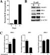
RBL2 mRNA and protein levels do not fluctuate in proliferating normal B cells. (A) To monitor normal B-cell activation and proliferation, the fraction of BrdU-positive cells was determined after 0, 2, and 4 days of treatment with recombinant human IL-4 and goat anti-human IgG + IgM. The percentage of BrdU incorporation was calculated by dividing the number of BrdU-positive cells by the total number of cells counted. (B) Whole-cell extracts were collected from resting (R) or activated (A) B cells 4 days after IL-4 and anti-human IgG + IgM treatment, and 25 μg of total protein was analyzed by Western blotting using the indicated antibodies. Anti-β-actin is included to show equal loading. (C) mRNA levels of the RB family of tumor suppressor genes were analyzed by real-time RT-PCR twice in triplicate, as described in the legend to Fig. 5A.
FIG. 7.
FIG. 7.
Reducing expression of PRMT5 impairs cancer cell growth. (A) To reduce PRMT5 protein expression, the transformed WaC3CD5 B-CLL cell line was infected with either recombinant lentivirus containing either vector alone or vector including AS-PRMT5 DNA. After 0, 2, and 4 days, 25 μg of whole-cell extracts was analyzed by Western blotting using anti-PRMT5 and anti-β-actin antibodies. (B) The growth rates of PRMT5 knockdown and control cell lines were analyzed every 2 days for 6 days. Shown is the average of three independent experiments. (C) Steady-state levels of RBL2 mRNA were analyzed after 0, 2, and 4 days by real-time RT-PCR using total RNA from control and AS-PRMT5 cells. (D) Whole-cell extracts (25 μg) were collected from control and AS-PRMT5 cells and subjected to Western blotting with anti-PRMT5, anti-RBL1, anti-RB1, anti-RBL2, and anti-β-actin antibodies.
FIG. 8.
FIG. 8.
siRNA-mediated knockdown of PRMT5 results in RBL2 derepression and the slow growth of transformed B cells. (A) Transformed WaC3CD5 B-CLL cells were treated either with control (Ctrl) siRNA, siPRMT5, or siPRMT5 combined with siRBL2, and 25 μg of whole-cell extracts was analyzed by Western blotting at the indicated times using anti-PRMT5, anti-RBL2, anti-RB1, and anti-RBL2 antibodies. Anti-β-actin served as a control. (B) The growth rate of transformed WaC3CD5 B-CLL cells electroporated with either Ctrl siRNA, siPRMT5, or siPRMT5 combined with siRBL2 by calculating the number of viable cells for 0, 2, and 4 days. Each data point represents the averages from three independent experiments. (C) Real-time RT-PCR was used to measure the steady-state levels of E2F1, RR2, and β-ACTIN using total RNA from WaC3CD5 cells electroporated with Ctrl siRNA, siPRMT5, or siPRMT5 and siRBL2 as described in the legend to Fig. 5A.
FIG. 9.
FIG. 9.
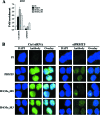
siRNA-mediated knockdown of PRMT5 abolishes gene-specific and global H3R8 and H4R3 symmetric methylation. (A) ChIP analysis was used to assess the association of PRMT5 with the RBL2 promoter and the level of H3R8 and H4R3 methylation in WaC3CD5 cells electroporated with either control (Ctrl) siRNA or siPRMT5. (B) To visualize the effect of PRMT5 knockdown on the global symmetric methylation of histones H3R8 and H4R3, immunofluorescence staining was conducted as described in the legend to Fig. 1B. WaC3CD5 cells were electroporated either with Ctrl siRNA or siPRMT5, and cells were stained after 4 days with either preimmune (PI), anti-PRMT5, anti-H3(Me2)R8, or anti-H4(Me2)R3 antibody. Pictures were taken at 100× magnification.

References

    1. Bartel, D. P. 2004. MicroRNAs: genomics, biogenesis, mechanism, and function. Cell 116281-297. - PubMed
    1. Bedford, M. T., and S. Richard. 2005. Arginine methylation an emerging regulator of protein function. Mol. Cell 18263-272. - PubMed
    1. Blais, A., C. J. van Oevelen, R. Margueron, D. Acosta-Alvear, and B. D. Dynlacht. 2007. Retinoblastoma tumor suppressor protein-dependent methylation of histone H3 lysine 27 is associated with irreversible cell cycle exit. J. Cell Biol. 1791399-1412. - PMC - PubMed
    1. Brehm, A., and T. Kouzarides. 1999. Retinoblastoma protein meets chromatin. Trends Biochem. Sci. 24142-145. - PubMed
    1. Chapman, E. J., and J. C. Carrington. 2007. Specialization and evolution of endogenous small RNA pathways. Nat. Rev. Genet. 8884-896. - PubMed

Publication types

MeSH terms