. 2008 Aug 11;182(3):437-47.
doi: 10.1083/jcb.200805124.

TRPP2 and TRPV4 form a polymodal sensory channel complex

Affiliations

TRPP2 and TRPV4 form a polymodal sensory channel complex

Michael Köttgen et al. J Cell Biol. .

Abstract

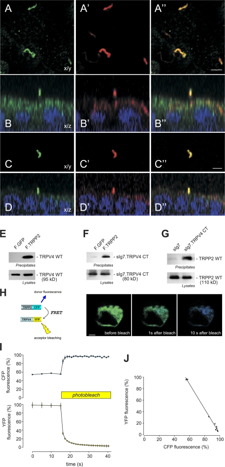
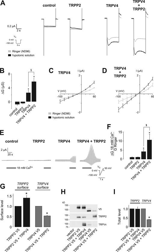
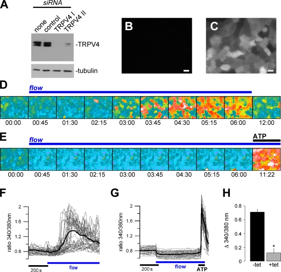
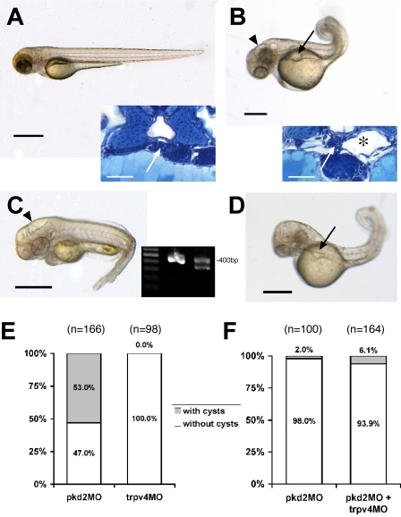
The primary cilium has evolved as a multifunctional cellular compartment that decorates most vertebrate cells. Cilia sense mechanical stimuli in various organs, but the molecular mechanisms that convert the deflection of cilia into intracellular calcium transients have remained elusive. Polycystin-2 (TRPP2), an ion channel mutated in polycystic kidney disease, is required for cilia-mediated calcium transients but lacks mechanosensitive properties. We find here that TRPP2 utilizes TRPV4 to form a mechano- and thermosensitive molecular sensor in the cilium. Depletion of TRPV4 in renal epithelial cells abolishes flow-induced calcium transients, demonstrating that TRPV4, like TRPP2, is an essential component of the ciliary mechanosensor. Because TRPV4-deficient zebrafish and mice lack renal cysts, our findings challenge the concept that defective ciliary flow sensing constitutes the fundamental mechanism of cystogenesis.


Figures

Figure 1.
Figure 1.
TRPV4 and TRPP2 interact and colocalize in primary cilia. (A) Subcellular localization of native TRPV4 and TRPP2 in polarized MDCK cells. TRPV4 localizes to primary cilia. Confocal images were acquired at the level of the apical membrane. TRPV4 (A) and acetylated tubulin (A′) colocalize in the primary cilium (A′′; merge). (B) Confocal z sections show that the primary cilium emerges from the apical membrane (B: anti-TRPV4; B′: anti-acetylated tubulin; B′′: merge).(C) TRPV4 and TRPP2 (C′) colocalize in the primary cilium (C′′; merge). (D) z section of a confocal image z stack of the cells shown in C. (E) Coimmunoprecipitation of TRPP2 and TRPV4 in HEK 293 cells. The flag-tagged carboxy terminus of TRPP2 (F.TRPP2) coprecipitates wild-type TRPV4, and the carboxy terminus of TRPV4 fused to a membrane-anchored immunoglobulin tag (sIg7.TRPV4 CT; F). (G) The same TRPV4 fusion protein precipitates TRPP2 wild type (WT). (H) FRET between TRPP2-CFP and TRPV4-YFP was revealed by increase in donor fluorescence after acceptor bleaching. HEK 293 cells were transfected with TRPP2-CFP and TRPV4-YFP. TRPP2-CFP was excited at 458 nm, and the emitted CFP and YFP fluorescence was recorded before and after photobleaching of the YFP fluorescence at 488 nm. (I) Time course of the normalized CFP and YFP fluorescence during photobleaching experiments (n = 5). (J) Correlation of the relative amount of YFP photobleaching and the concomitant increase in CFP fluorescence in the same cell. Bars, 5 μm.
Figure 2.
Figure 2.
Functional interaction of TRPP2 and TRPV4 in X. laevis oocytes. (A) Analysis of TRP channel whole-cell currents under voltage clamp (Vc) conditions. Currents were recorded in X. laevis oocytes injected with cRNA encoding TRPP2 and/or TRPV4. Mean currents in Ringer or hypotonic solution are shown. (B) Summary of data acquired in A. Asterisks indicate significant differences in the hypotonicity-induced whole-cell conductance (ΔG) compared with water-injected control oocytes; §, significant difference between bars as indicated (n = 21, 15, 38, and 37, respectively). (C and D) Current-voltage (I–V) relations for oocytes expressing TRPV4 or TRPV4 and TRPP2 (D; n = 7). (E) Increasing the extracellular Ca2+ concentration from 1.8 to 18 mM led to a significant increase in whole-cell currents in TRPV4-expressing oocytes. This effect was dramatically augmented in oocytes coexpressing TRPP2. Currents were continuously monitored under voltage clamp conditions (Vc protocol as indicated). (F) Analysis of the relative Ca2+ conductance revealed that TRPP2 significantly increased the Ca2+ currents (normalized group data, GCa2+/GRinger from E; n = 10, 10, 11, and 7, respectively). Whole-cell currents of oocytes expressing TRPV4 with or without TRPP2 were inhibited by RR with similar potency (see Fig. S2, available at http://www.jcb.org/cgi/content/full/jcb.200805124/DC1). (G) Analysis of the surface expression of TRPP2 and TRPV4 (n = 4) using an enzyme-linked assay for detection of both channels at the plasma membrane. Asterisks indicate significant differences between black and grey bars, respectively. (H) Representative Western blot of total protein amount of a representative experiment. (I) Summarized data of total protein amounts (n = 4). Error bars represent mean values ± SEM.
Figure 3.
Figure 3.
TRPV4 is required for flow-mediated Ca2+ signals in renal epithelial cells. (A) HEK 293 cells overexpressing V5-tagged canine TRPV4 were transiently transfected with empty vector (none), an ineffective siRNA construct (control), or two different effective siRNA constructs. TRPV4 expression levels were monitored by Western blotting against V5 (with tubulin as a loading control). (B and C) MDCK cells stably expressing tetracycline-inducible TRPV4 II siRNA. siRNA expression in the absence (B) and presence of tetracycline (C) can be monitored by concomitant expression of GFP. (D) Flow-mediated Ca2+ signaling was studied in ciliated MDCK cells using Fura-2. The time course of cytosolic Ca2+ increase in response to laminar flow (ciliary bending) is shown in representative Fura-2 pseudocolor images reflecting the Ca2+ concentration in MDCK cells (340/380 nm fluorescence ratio; blue/green: low [Ca2+]c; yellow/red: high [Ca2+]c; time is indicated in min:s). (E) Flow-induced Ca2+ signals in cells expressing TRPV4 siRNA. Although the flow-induced response was abolished upon knockdown of TRPV4, 10 μM ATP still elicited a robust Ca2+ increase. (F) Single-cell analysis of the Ca2+ signals (ratio 340/380 nm) of the same experiment as shown in D (each trace represents an individual cell and the bold line depicts the mean; for better visibility, only 50% of cells in the visual field are represented). (G) Effect of TRPV4 siRNA on flow-induced Ca2+ signals. Single-cell analysis of the same experiment as shown in E; again, 50% of cells in the visual field are represented. (H) Grouped data from three independent experiments (n = 3; −tet: 75 cells per n; +tet: 93 cells per n). All cells in the visual field were included to calculate the flow-induced Ca2+ peak baseline. *, statistical significance. Error bars represent mean values ± SEM. Bars, 10 μm.
Figure 4.
Figure 4.
Role of TRPP2 and TRPV4 in the pathogenesis of pronephric cysts in zebrafish. (A) Wild-type larva (55 hpf) with histologically normal glomerulus (inset, arrow) and adjacent tubules. Bars, 500 μm. (B) Disruption of TRPP2 function in pkd2 morpholino (pkd2MO)-injected larva (55 hpf) results in dorsally flexed curly tail, hydrocephalus (arrowhead), and pronephric cyst formation (arrow), which is confirmed histologically. The pronephric tubules are dilated (inset, *) and the glomerulus is stretched (inset, arrow). (C) trpv4 morphant larva (55 hpf) show hydrocephalus (arrowhead) but lack pronephric cysts (grouped data are shown in E). The effect of the trpv4 splice morpholino was verified by RT-PCR (inset) from single embryo total RNA (55 hpf), with nested primers in flanking exons yielding a 400-bp amplicon in wild-type embryos (middle lane) and an additional shorter amplicon in the morphant embryo (right lane; 100 bp marker, left lane). Sequencing revealed an in-frame deletion of the whole seventh coding exon (93 bp) and therefore the loss of most of the second transmembrane domain. Higher trpv4MO doses led to substantial increase of lethality in the injected embryos. To examine an interaction between the two proteins with respect to cyst formation, the embryos were injected with both morpholinos. (D) With two morpholinos, pkd2MO and trpv4MO, the coinjected embryo (55 hpf) shows a pronephric cyst (arrow). For the coinjection, the dose of the pkd2MO was titrated to levels at which the occurrence of cysts is very low, and trpv4MO was added in a medium dose with low lethality. The additive effect showed no significant increase in incidence of cysts, as is shown in the bar graph (F).
Figure 5.
Figure 5.
TRPP2 and TRPV4 form a thermosensory complex in vitro and in vivo. (A) Analysis of TRP channel whole-cell currents under voltage clamp (Vc) conditions. Currents were recorded in X. laevis oocytes injected with cRNA encoding TRPP2 and/or TRPV4. Representative inward currents at 20°C or 39°C are shown. (B) Summary of data acquired in A. Asterisks indicate significant differences in the temperature-activated whole cell conductance (ΔG) compared with water-injected control oocytes; §, significant differences between data as indicated (n = 4, 4, 4, and 6, respectively). (C) Current-voltage (I–V) relations for oocytes expressing TRPV4 or TRPV4 and TRPP2 (D; gray: 20°C; black: 39°C). (E) Tail withdrawal latencies after immersion into a water bath at moderately hot temperatures were measured in mice of the indicated genotypes (n = 10 per genotype; asterisk indicates significant difference compared with wild-type [WT] and TRPV4+/− mice; §, significant difference from TRPP2+/− mice). (F) Tail withdrawal latencies at noxiously hot temperatures (n = 10 per genotype). Error bars represent mean values ± SEM.

References

    1. Arnould, T., L. Sellin, T. Benzing, L. Tsiokas, H.T. Cohen, E. Kim, and G. Walz. 1999. Cellular activation triggered by the autosomal dominant polycystic kidney disease gene product PKD2. Mol. Cell. Biol. 19:3423–3434. - PMC - PubMed
    1. Cuajungco, M.P., C. Grimm, K. Oshima, D. D'Hoedt, B. Nilius, A.R. Mensenkamp, R.J. Bindels, M. Plomann, and S. Heller. 2006. PACSINs bind to the TRPV4 cation channel. PACSIN 3 modulates the subcellular localization of TRPV4. J. Biol. Chem. 281:18753–18762. - PubMed
    1. Giamarchi, A., F. Padilla, B. Coste, M. Raoux, M. Crest, E. Honore, and P. Delmas. 2006. The versatile nature of the calcium-permeable cation channel TRPP2. EMBO Rep. 7:787–793. - PMC - PubMed
    1. Gradilone, S.A., A.I. Masyuk, P.L. Splinter, J.M. Banales, B.Q. Huang, P.S. Tietz, T.V. Masyuk, and N.F. Larusso. 2007. Cholangiocyte cilia express TRPV4 and detect changes in luminal tonicity inducing bicarbonate secretion. Proc. Natl. Acad. Sci. USA. 104:19138–19143. - PMC - PubMed
    1. Guler, A.D., H. Lee, T. Iida, I. Shimizu, M. Tominaga, and M. Caterina. 2002. Heat-evoked activation of the ion channel, TRPV4. J. Neurosci. 22:6408–6414. - PMC - PubMed

Publication types

Substances