. 2008 Aug 11;182(3):509-17.
doi: 10.1083/jcb.200801079.

A cyclin D1/microRNA 17/20 regulatory feedback loop in control of breast cancer cell proliferation

Affiliations

A cyclin D1/microRNA 17/20 regulatory feedback loop in control of breast cancer cell proliferation

Zuoren Yu et al. J Cell Biol. .

Abstract

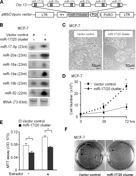
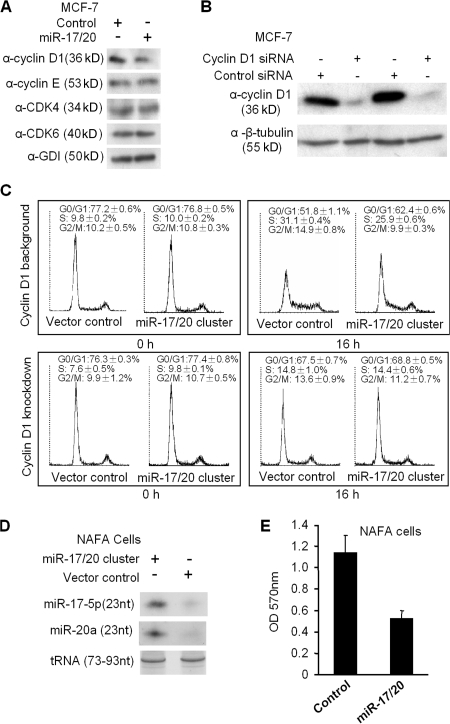
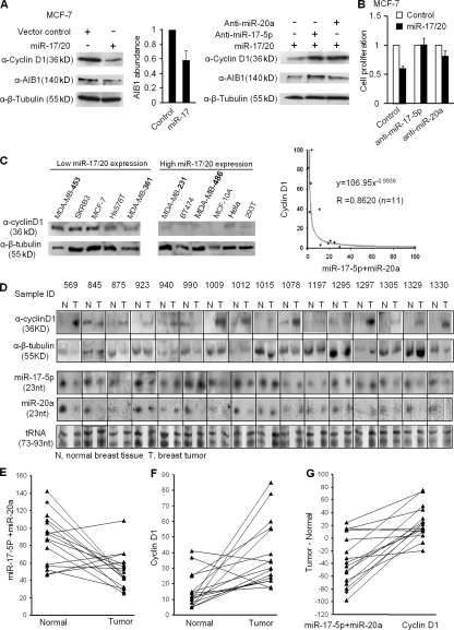
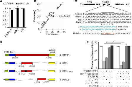
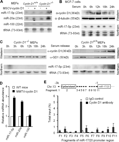
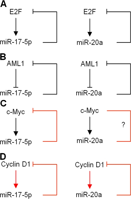
Decreased expression of specific microRNAs (miRNAs) occurs in human tumors, which suggests a function for miRNAs in tumor suppression. Herein, levels of the miR-17-5p/miR-20a miRNA cluster were inversely correlated to cyclin D1 abundance in human breast tumors and cell lines. MiR-17/20 suppressed breast cancer cell proliferation and tumor colony formation by negatively regulating cyclin D1 translation via a conserved 3' untranslated region miRNA-binding site, thereby inhibiting serum-induced S phase entry. The cell cycle effect of miR-17/20 was abrogated by cyclin D1 siRNA and in cyclin D1-deficient breast cancer cells. Mammary epithelial cell-targeted cyclin D1 expression induced miR-17-5p and miR-20a expression in vivo, and cyclin D1 bound the miR-17/20 cluster promoter regulatory region. In summary, these studies identify a novel cyclin D1/miR-17/20 regulatory feedback loop through which cyclin D1 induces miR-17-5p/miR-20a. In turn, miR-17/20 limits the proliferative function of cyclin D1, thus linking expression of a specific miRNA cluster to the regulation of oncogenesis.


Figures

Figure 1.
Figure 1.
miR-17/20 inhibits breast cancer cell proliferation. (A) The genomic region of miR-17/20 cluster on chromosome 13q31 and the vector structure for miRNA17/20 overexpression. (B) Northern blot analysis demonstrating the increased expression of the six members of miR-17/20 cluster in the miR-17/20–transduced MCF-7 cells. tRNA served as loading control. (C and D) Cellular proliferation assay; (E) an MTT assay; and (F) a colony formation assay consistently showed the inhibition of MCF-7 cell proliferation by miR-17/20 overexpression. Cellular proliferation assays and the MTT assay were performed in three independent experiments (data are equal to mean ± SEM). *, P < 0.01.
Figure 2.
Figure 2.
miR-17/20 arrests cell cycle at the G1 phase in a cyclin D1–dependent manner. (A) Western blot analysis of the cyclin D1, CDK4, CDK6, and cyclin E expression in miR-17/20–transduced cells. GDI served as loading control. (B) MCF-7 cells were transfected with cyclin D1 siRNA and control siRNA. Western blotting demonstrated the efficient knockdown of cyclin D1 after 72 h of siRNA treatment. (C) Cell cycle analysis indicated the increased population of cells at G0/G1 phase and decreased S and G2/M phase cells in miR-17/20–transduced MCF-7 cells under a cyclin D1 background. This difference was abolished by knockdown of cyclin D1 in cells. The analysis was performed in triplicates (data are equal to mean ± SEM). (D) Northern blot demonstrated the increased expression of miR-17/20 in the miR-17/20–transduced NAFA cells. tRNA served as a loading control. (E) The MTT assay showed the inhibited cell proliferation of NAFA cells by miR-17/20 transduction. The assay was performed in three independent experiments, and the data are presented as the mean ± SEM.
Figure 3.
Figure 3.
The inverse correlation between cyclin D1 abundance and miR-17/20 expression in human breast cancer tissues and cell lines. (A) Western blots showed the repression of cyclin D1 and AIB1 abundance by miR-17/20. Both anti–miR-17-5p and anti–miR-20a increased the expression of cyclin D1 and AIB1 in cells. AIB1 is a positive control. β-tubulin was a loading control. (B) Cotransfection of anti–miR-17-5p or anti–miR-20a reversed the antiproliferative function of miR-17/20 in MCF-7 cells. (C) Western blots showed the high cyclin D1 level in low miR-17/20–expressing breast cancer cell lines, and low cyclin D1 in high miR-17/20 cell lines. A Northern blot of miRNA17/20 is shown in Fig. S1 (available at http://www.jcb.org/cgi/content/full/jcb.200801079/DC1). (D) The abundances of cyclin D1 and miR-17-5p/miR-20a in 16 human breast tumor tissues and 16 matching normal breast tissues were determined by Western and Northern blots. Tissue sample IDs were provided by the sample provider. Black lines indicate that intervening lanes have been spliced out. N, normal tissue; T, tumor tissue. (E) Statistically significant down-regulation of miR-17/20 expression in breast tumors over matching normal tissue. P = 0.004 by Wilcoxon signed rank test. The grayscale intensity of each band in D was obtained by AlphaImager software. The y axis value stands for the addition of miR-17-5p and miR-20a expression in each sample. (F) Significant up-regulation of cyclin D1 expression in breast tumors over matching normal tissue. P = 0.001. (G) Plotting the paired difference of tumor and normal samples expression for each marker (miR-17/20 vs. cyclin D1). The exact McNemar's test indicates a significant association between the up-regulation of expression in cyclin D1 and the down-regulation of miR-17/20 expression. P = 0.002.
Figure 4.
Figure 4.
miR-17/20 represses cyclin D1 expression through a conserved 3′ UTR site. (A) Quantitative real-time RT-PCR assay did not show significant difference in mRNA level of cyclin D1 between miR-17/20–transduced cells and control cells. ABI1 was a positive control. 18S served as an internal control for normalization. This experiment was repeated three times in triplicate. Data are mean ± SEM. (B) BLASTN analysis of human and mouse cyclin D1 mRNAs identified a miR-17-5p– and miR-20a–binding site at the conserved 3′ UTR region. (C) Sequence alignment of the miR-17-5p and miR-20a base-paring site in the 3′ UTR of cyclin D1 mRNAs. The region complementary to the 2–10 nt of miR-17/20 is highly conserved among human, mouse, rat, and dog. The “seed” sequence of miR-17/20 that is complementary to cyclin D1 is shown in italics and boxed. The mutant sequence is identical to the wild-type sequence except the mutated nucleotides are shown in red. (D) Luciferase reporter assay constructs. 3′ UTR FL, the full-length 3′ UTR of cyclin D1 inserted to the downstream of the luciferase coding region in the pGL3 vector; 3′ UTR-1, a fragment of cyclin D1 3′ UTR containing the miR-17/20–binding sequence; 3′ UTR-1-mu, the mutated construct identical to 3′UTR-1 but point mutated in the miR-17/20 binding site; 3′ UTR-2, another fragment of cyclin D1 3′ UTR without the miR-17/20–binding sequence. (E) Luciferase reporter assay showed the decreased luciferase activity in miR-17/20 overexpressed cells for both 3′ UTR FL and 3′ UTR-1 constructs, but not for the PGL-3 empty vector, 3′UTR-1-mu and 3′UTR-2 constructs. The luciferase activity was normalized to β-galactosidase. Data are derived from three independent experiments. Values are presented as the mean ± SEM (n = 3). *, P < 0.01
Figure 5.
Figure 5.
Cyclin D1 induces miR-17-5p and miR-20a expression. (A) Northern blotting showed the increased expression of miR-17-5p and miR-20a in the MSCV–cyclin D1–infected MEF cells. miR-100 was used as a negative control. tRNA served as loading control. (B) Breast cancer cell line MCF-7. (C) cyclin D1−/−, and cyclin D1+/+ MEFs were starved by 5% charcoal-stripped serum for 48 h, followed by 10% FBS stimulation. The time course examination of the cyclin D1 protein level and the miR-17-5p expression level were performed by Western and Northern blots, respectively. The results showed the induction of cyclin D1 and miR-17-5p by serum stimulation in the MCF-7 and cyclin D1+/+ MEF cells, but not in cyclin D1−/− MEFs. (D) miR-17-5p and miR-20a detection in the MMTV–cyclin D1 transgene induced mammary tumors (n = 3) compared with normal mice mammary gland. Data from three mammary tumor samples were presented as mean ± SEM. (E) Real-time PCR analysis of cyclin D1 chromatin immunoprecipitated DNA. 11 fragments derived from chromosome 11q31 upstream of miR-17/20 cluster were examined. For each fragment, amplifications on chromatin before immunoprecipitation and chromatin immunoprecipitated with preimmune serum were performed as input and negative control, respectively. This experiment was repeated three times; data are equal to mean ± SEM.
Figure 6.
Figure 6.
miR-17/20 regulatory loops. (A) E2F-miR-17/20 regulatory loop. (B) AML1-miR-17/20 regulatory loop. (C) c-Myc–miR-17/20 regulatory loop. (D) cyclin D1–miR-17/20 regulatory loop. Red arrows are supported by our data. Black arrows are supported by scientific literature.

References

    1. Albanese, C., J. Johnson, G. Watanabe, N. Eklund, D. Vu, A. Arnold, and R.G. Pestell. 1995. Transforming p21ras mutants and c-Ets-2 activate the cyclin D1 promoter through distinguishable regions. J. Biol. Chem. 270:23589–23597. - PubMed
    1. Ambros, V. 2004. The functions of animal microRNAs. Nature. 431:350–355. - PubMed
    1. Calin, G.A., and C.M. Croce. 2006. MicroRNA signatures in human cancers. Nat. Rev. Cancer. 6:857–866. - PubMed
    1. Calin, G.A., C.D. Dumitru, M. Shimizu, R. Bichi, S. Zupo, E. Noch, H. Aldler, S. Rattan, M. Keating, K. Rai, et al. 2002. Frequent deletions and down-regulation of micro- RNA genes miR15 and miR16 at 13q14 in chronic lymphocytic leukemia. Proc. Natl. Acad. Sci. USA. 99:15524–15529. - PMC - PubMed
    1. Calin, G.A., C. Sevignani, C.D. Dumitru, T. Hyslop, E. Noch, S. Yendamuri, M. Shimizu, S. Rattan, F. Bullrich, M. Negrini, and C.M. Croce. 2004. Human microRNA genes are frequently located at fragile sites and genomic regions involved in cancers. Proc. Natl. Acad. Sci. USA. 101:2999–3004. - PMC - PubMed

Publication types