. 2008 Aug 11;182(3):531-42.
doi: 10.1083/jcb.200711151.

Regulation of protein O-glycosylation by the endoplasmic reticulum-localized molecular chaperone Cosmc

Affiliations

Regulation of protein O-glycosylation by the endoplasmic reticulum-localized molecular chaperone Cosmc

Tongzhong Ju et al. J Cell Biol. .

Abstract

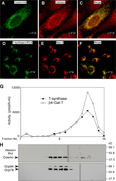
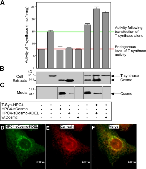
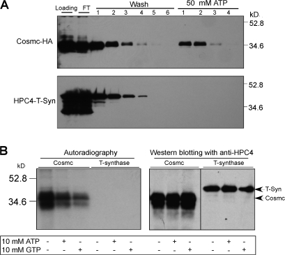
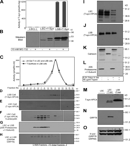
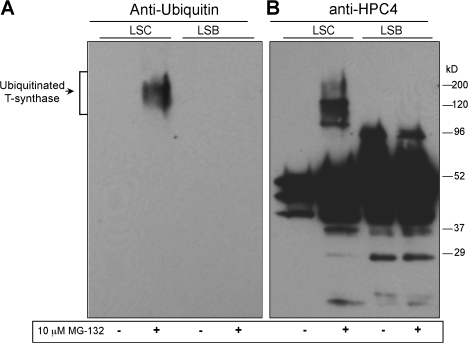
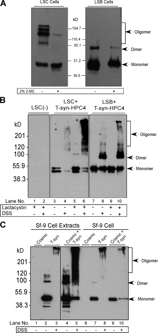
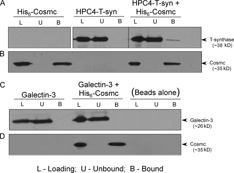
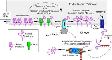
Regulatory pathways for protein glycosylation are poorly understood, but expression of branchpoint enzymes is critical. A key branchpoint enzyme is the T-synthase, which directs synthesis of the common core 1 O-glycan structure (T-antigen), the precursor structure for most mucin-type O-glycans in a wide variety of glycoproteins. Formation of active T-synthase, which resides in the Golgi apparatus, requires a unique molecular chaperone, Cosmc, encoded on Xq24. Cosmc is the only molecular chaperone known to be lost through somatic acquired mutations in cells. We show that Cosmc is an endoplasmic reticulum (ER)-localized adenosine triphosphate binding chaperone that binds directly to human T-synthase. Cosmc prevents the aggregation and ubiquitin-mediated degradation of the T-synthase. These results demonstrate that Cosmc is a molecular chaperone in the ER required for this branchpoint glycosyltransferase function and show that expression of the disease-related Tn antigen can result from deregulation or loss of Cosmc function.


Figures

Figure 1.
Figure 1.
Localization of human Cosmc and T-synthase. (A–F) Immunofluorescent staining. CHO K1 cells cultured on chambered slides were transiently transfected with Cosmc-HA or with T-synthase–HPC4 and stained with rat anti-HA IgG1 (green) and rabbit anti-calnexin IgG (red; A–C; bars, 4 μm) or with mouse anti-HPC4 (green) and rabbit anti–α-ManII (red; D–F; bars, 8 μm). (G and H) Sucrose gradient subcellular fractionation. 293T cells transiently transfected with HPC4-Cosmc were harvested and homogenized. The postnuclear supernatant (PNS) was loaded onto a sucrose gradient. After ultracentrifugation, 16 fractions (∼0.6 ml/fraction) were collected and measured for both T-synthase and β4–Gal-T activity (G) and analyzed on Western Blot with anti-HPC4 and anti-KDEL (H).
Figure 2.
Figure 2.
KDEL-tagged soluble form of Cosmc functions as wtCosmc and localizes in ER. (A–C) Function of KDEL-tagged soluble form of Cosmc. 293T cells were transiently transfected with plasmids encoding T-synthase–HPC4, HPC4-sCosmc, HPC4-sCosmc-KDEL, and wtCosmc. Cell extracts were prepared and T-synthase activity was measured. Bar 1 represents a single value, bar 5 represents a mean of duplicate values with error bar shown, and the remaining bars represent a mean of triplicate values with error bars shown. The pink line indicates the endogenous level of T-synthase activity and the green line represents the activity after transfection of T-synthase alone (A). T-synthase and Cosmc in cell extracts and media were also analyzed by Western blot with anti-HPC4 (B and C). Black lines indicate that intervening lanes have been spliced out. (D–F) Localization of sCosmc-KDEL. CHO K1 cells cultured on chambered slides transiently transfected with HPC4-sCosmc-KDEL were immunofluorescently stained with anti-HPC4 (green; D) and rabbit anti-calnexin IgG (red; E) and merged (F). The images were collected by confocal microscopy. Bars, 8 μm.
Figure 3.
Figure 3.
Cosmc has ATP-binding activity. (A) Hi-5 insect cells were infected with Baculovirus encoding either Cosmc-HA or HPC4-sT-synthase plus wild-type Cosmc. Cells for Cosmc-HA and media from cell coexpressing HPC4–sT-synthase and wild-type Cosmc were harvested. The cell extracts and media were loaded on ATP-Sepharose column for chromatography and eluted with 50 mM ATP, respectively. The washes and eluates were analyzed by Western blot with anti-HA for Cosmc and anti-HPC4 for T-synthase, as indicated. (B) HPC4-sCosmc and HPC4–sT-synthase (coexpressed with wtCosmc) were expressed in Hi-5 cells and purified from the media. 3 μg of Cosmc and T-synthase were photolabeled with 8-Azido α-[32P]ATP, analyzed on SDS-PAGE, and transferred to nitrocellulose membrane for radioautogram and Western blot with anti-HPC4. Black line indicates that intervening lanes have been spliced out.
Figure 4.
Figure 4.
Characterization of T-synthase expressed in LSC cells containing a dysfunctional Cosmc. (A and B) MG-132 causes accumulation of full-length T-synthase protein in LSC cells but failed to restore its activity. LSC and LSB cells stably expressing T-synthase–HPC4 were treated with 10 μM MG-132 for 12 h and harvested. Cell extract was made and one portion was used for measuring T-synthase activity (A), whereas the other portion was analyzed on SDS-PAGE (30 μg protein/lane) by Western blot with anti-HPC4 (B). Error bars represent ±1 SD from the mean. Black lines indicates that intervening lanes have been spliced out. (C–H) T-synthase expressed in LSC cells resides mainly in heavy membrane fractions (RER), whereas T-synthase from LSB cells is in light membrane fractions (Golgi). LSC and LSB cells as in A and B treated with 10 μM lactacystin were homogenized and cell homogenate was fractionated on a sucrose gradient by ultracentrifugation. The fractions were collected and the activities of β4–Gal-T and T-synthase in fractions were determined (C). The fractions were also analyzed by Western blotting with anti-HPC4 (D–F), anti-20S proteasome α1-subunit (G), and anti-KDEL (GRP78 and GRP94; H). In F, lane 2 represents the combination of fractions 1 and 2. (I–O) T-synthase expressed in LSC cells resides mainly in the lumen of the RER and was associated with GRP78. The cell PNS of LSC and LSB cells as in A and B was digested with 40 μg/ml trypsin in the presence or absence of 0.2% Triton X-100, and T-synthase was analyzed under reducing SDS-PAGE by Western blotting with HPC4 mAb (I and J). As controls, calnexin and 20S proteasome α1-subunit were also analyzed by Western blotting with their respective antibody (K and L). T-synthase was purified from LSC and LSB cells and Western blotted with anti-HPC4 (M) and anti-KDEL (GRP78; N). GRP78 in the cell extract corresponding to one-tenth of the material was detected to confirm comparable starting amounts of material (O).
Figure 5.
Figure 5.
T-synthase expressed in LSC Cells is partially ubiquitinated. LSC and LSB cells stably expressing T-synthase–HPC4 were treated with MG-132 for 12 h and subsequently harvested. T-synthase from 200 μl of cell extract (500 μg of protein total) was purified and analyzed by Western blot with anti-ubiquitin (A) first, and then with anti-HPC4 (B) after stripping of the membrane.
Figure 6.
Figure 6.
T-synthase from cells in the absence of a functional Cosmc is mainly in covalent oligomers. T-synthase–HPC4 expressed in LSC and LSB cells treated with 10 μM lactacystin was affinity purified and analyzed on SDS-PAGE under reducing and nonreducing conditions and Western blotted with anti-HPC4 (A). Cell extracts were made and cross-linking was performed by adding 5 mM DSS. The recombinant T-synthase was purified and analyzed by Western blotting under reducing conditions (B). Sf-9 cells infected with Baculovirus-expressing T-synthase-HPC4 or coinfected with Baculovirus encoding wtCosmc were harvested and one portion was made for cell extracts. The cell extracts and the other portion of intact cells were treated with DSS. T-synthase was purified from the cell extracts and analyzed by Western blot with anti-HPC4 (C). Black line indicates that intervening lanes have been spliced out.
Figure 7.
Figure 7.
Cosmc directly interacts with T-synthase. The N-terminal His-tagged soluble Cosmc were expressed in Hi-5 insect cells. The human N-terminal HPC4 epitope-tagged soluble T-synthase was expressed by cotransfecting WT Cosmc in Hi-5 insect cells. Proteins were purified directly from the media and used in coprecipitation experiments to test physical association. Galectin-3 was used as a negative control for specificity. Cosmc, T-synthase, Cosmc and T-Synthase, Galectin-3, Cosmc and Galactin-3 were incubated with Ni-NTA Superflow. Ni-NTA Superflow beads were washed and proteins were eluted and analyzed by Western blot with mAb to the HPC4, anti-Galectin-3, and anti-Cosmc. Cosmc interactions with T-synthase were determined (A and B). As a control, Cosmc and Galectin-3 interactions were determined (C and D). Black line indicates that intervening lanes have been spliced out. L, loading; U, unbound; B, bound.
Figure 8.
Figure 8.
Working model for Cosmc function. Human Cosmc is localized predominantly in the ER where it interacts with the nascent polypeptide of human T-synthase. Cosmc, along with other ER chaperones such as HSP70 (BiP)/HSP40 and protein disulfide isomerase (PDI), assists its folding properly. Native T-synthase, which occurs mainly as a homodimer, exits to the Golgi apparatus, where it functions in synthesizing core 1 O-glycans (T-antigen). When Cosmc is mutated and dysfunctional, the nascent polypeptides of T-synthase form inactive aggregates or oligomers, which are associated with GRP78 (BiP), and subsequently proceed to the ERAD pathway where they are retrotranslocated from the ER to the cytosol, ubiquitinated, and subsequently degraded by the proteasome.

References

    1. Ahner, A., and J.L. Brodsky. 2004. Checkpoints in ER-associated degradation: excuse me, which way to the proteasome? Trends Cell Biol. 14:474–478. - PubMed
    1. Allen, A.C., P.S. Topham, S.J. Harper, and J. Feehally. 1997. Leucocyte beta 1,3 galactosyltransferase activity in IgA nephropathy. Nephrol. Dial. Transplant. 12:701–706. - PubMed
    1. An, G., B. Wei, B. Xia, J.M. McDaniel, T. Ju, R.D. Cummings, J. Braun, and L. Xia. 2007. Increased susceptibility to colitis and colorectal tumors in mice lacking core 3–derived O-glycans. J. Exp. Med. 204:1417–1429. - PMC - PubMed
    1. Berger, E.G. 1999. Tn-syndrome. Biochim. Biophys. Acta. 1455:255–268. - PubMed
    1. Bergeron, J.J., M.B. Brenner, D.Y. Thomas, and D.B. Williams. 1994. Calnexin: a membrane-bound chaperone of the endoplasmic reticulum. Trends Biochem. Sci. 19:124–128. - PubMed

Publication types

MeSH terms