

Brain morphology in older African Americans, Caribbean Hispanics, and whites from northern Manhattan
- PMID: 18695055
- PMCID: PMC2692286
- DOI: 10.1001/archneur.65.8.1053
Brain morphology in older African Americans, Caribbean Hispanics, and whites from northern Manhattan
Abstract
Background: Aging is accompanied by a decrease in brain volume and by an increase in cerebrovascular disease.
Objective: To examine the effects of age, sex, race/ethnicity, and vascular disease history on measures of brain morphology, including relative brain volume, ventricular volume, hippocampus and entorhinal cortex volumes, and white matter hyperintensity (WMH) burden, in a large community-based cohort of racially/ethnically diverse older adults without dementia.
Design: The associations of age, sex, race/ethnicity, and self-reported vascular disease history with brain morphology were examined in a cross-sectional study using multiple linear regression analyses. Sex x race/ethnicity interactions were also considered.
Setting: The Washington Heights-Inwood Columbia Aging Project, a community-based epidemiological study of older adults from 3 racial/ethnic groups (white, Hispanic, and African American) from northern Manhattan.
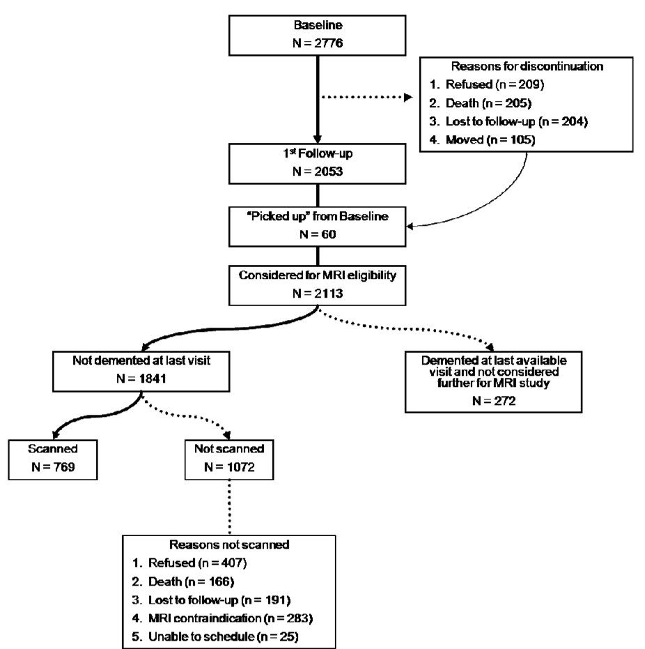
Participants: Beginning in 2003, high-resolution quantitative magnetic resonance (MR) images were acquired in 769 participants without dementia.
Main outcome measures: Relative brain volume (total brain volume/intracranial volume), ventricular volume, and hippocampus and entorhinal cortex volumes were derived manually on high-resolution MR images. White matter hyperintensities were quantified semiautomatically on fluid-attenuated inversion recovery-T2-weighted MR images.
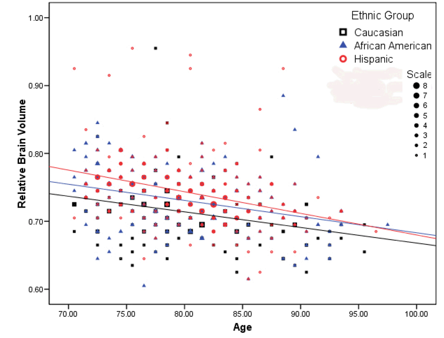
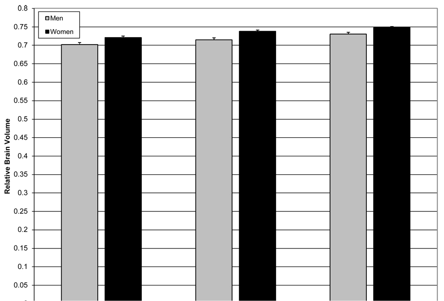
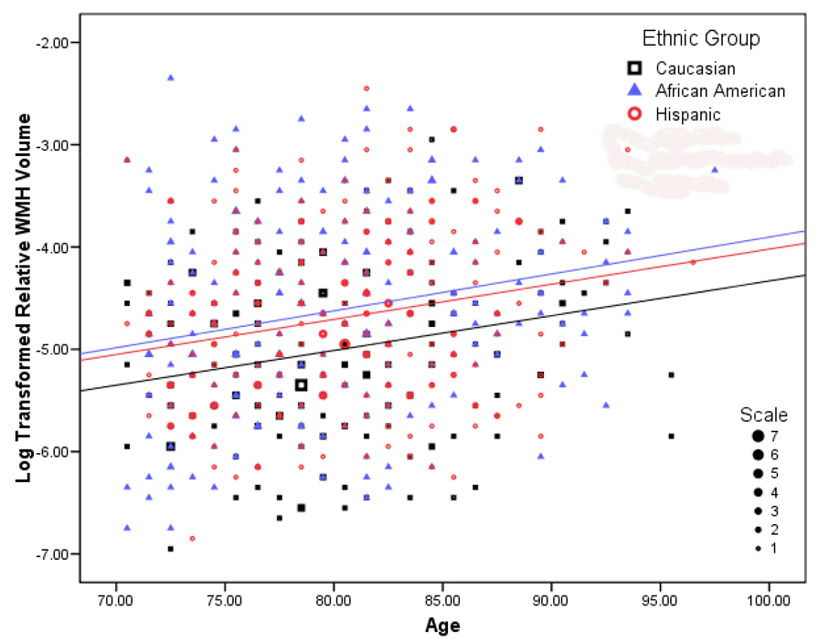
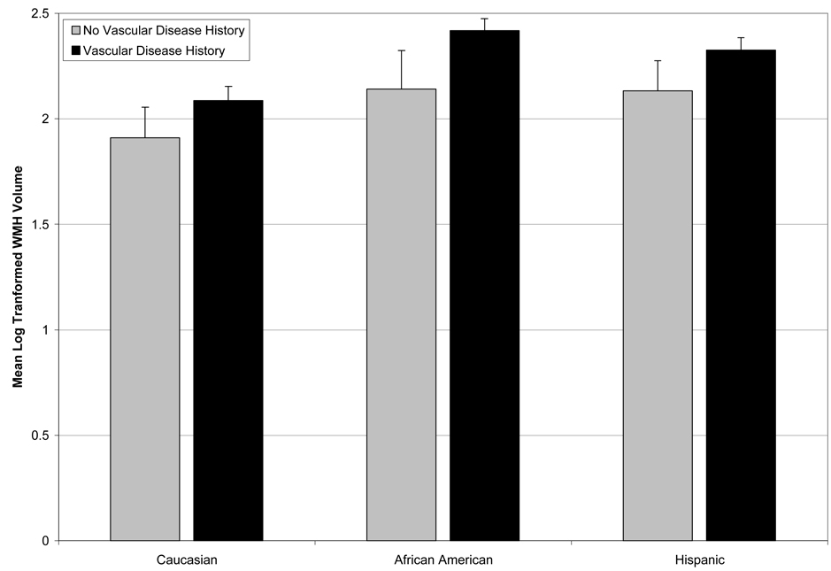
Results: Older age was associated with decreased relative brain volume and with increased ventricular and WMH volumes. Hispanic and African American participants had larger relative brain volumes and more severe WMH burden than white participants, but the associations of these variables with age were similar across racial/ethnic groups. Compared with men, women had larger relative brain volumes. Vascular disease was associated with smaller relative brain volume and with higher WMH burden, particularly among African Americans.
Conclusions: Older age and vascular disease, particularly among African Americans, are associated with increased brain atrophy and WMH burden. African American and Hispanic subjects have larger relative brain volumes and more WMH than white subjects. Racial/ethnic group differences in WMH severity seem to be partially attributable to differences in vascular disease. Future work will focus on the determinants and cognitive correlates of these differences.
Figures
References
-
- Raz N, Rodrigue KM, Kennedy KM, Acker JD. Vascular health and longitudinal changes in brain and cognition in middle-aged and older adults. Neuropsychology. 2007 Mar;21(2):149–157. - PubMed
-
- Courchesne E, Chisum HJ, Townsend J, et al. Normal brain development and aging: quantitative analysis at in vivo MR imaging in healthy volunteers. Radiology. 2000 Sep;216(3):672–682. - PubMed
-
- Tang Y, Whitman GT, Lopez I, Baloh RW. Brain volume changes on longitudinal magnetic resonance imaging in normal older people. J Neuroimaging. 2001 Oct;11(4):393–400. - PubMed
-
- Good CD, Johnsrude IS, Ashburner J, Henson RN, Friston KJ, Frackowiak RS. A voxel-based morphometric study of ageing in 465 normal adult human brains. Neuroimage. 2001 Jul;14(1 Pt 1):21–36. - PubMed
-
- Resnick SM, Goldszal AF, Davatzikos C, et al. One-year age changes in MRI brain volumes in older adults. Cereb Cortex. 2000 May;10(5):464–472. - PubMed
Publication types
MeSH terms
Grants and funding
LinkOut - more resources
Full Text Sources
Medical
