Case Reports
. 2008 Aug;126(8):1059-66.
doi: 10.1001/archopht.126.8.1059.

Temporary sutureless amniotic membrane patch for acute alkaline burns

Affiliations
Case Reports

Temporary sutureless amniotic membrane patch for acute alkaline burns

Ahmad Kheirkhah et al. Arch Ophthalmol. 2008 Aug.

Abstract

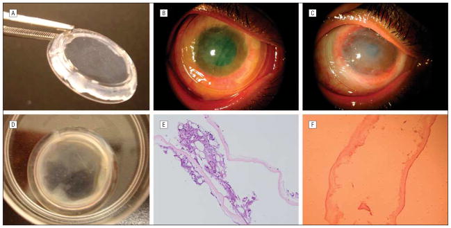
Objective: To evaluate the clinical outcome of a new sutureless approach for a temporary amniotic membrane patch (ProKera; Bio-Tissue, Inc, Miami, Florida) in eyes with acute burns.

Methods: Retrospective review of 5 eyes of 5 patients with grades I to III acute alkaline burns, receiving ProKera insertion within 8 days of injury.

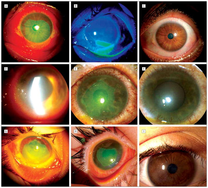
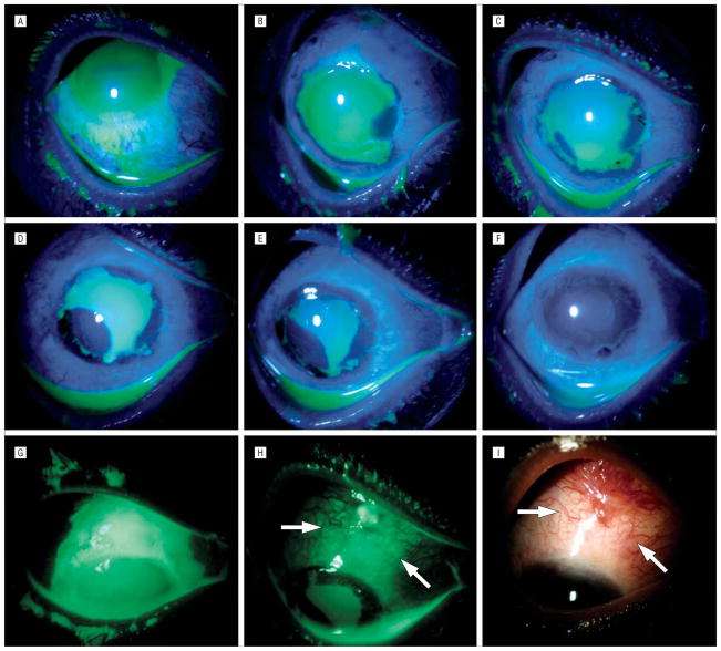
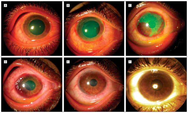
Results: These eyes had either total (2 cases) or extensive (60%-75%, 3 cases) corneal epithelial defects with limbal (120 degrees -360 degrees ) and conjunctival (30%-60%) epithelial defects. ProKera was inserted within a mean (SD) of 3.7 (3.1) days after burn and repeated 1 to 3 times for 3 cases. Conjunctival defects reepithelialized in 8.2 (5) days (range, 5-17 days), while limbal and corneal defects healed in 13.6 (8.3) days (range, 5-25 days). The latter was completed with circumferential closure of limbal defects followed by centripetal healing of corneal defects. In 3 eyes, early peripheral corneal neovascularization was followed by marked regression on completion of healing. During 16.8 (10.8) months of follow-up, all eyes retained a stable surface with improved corneal clarity, and without limbal deficiency or symblepharon.

Conclusion: This sutureless application of an amniotic membrane patch allows for early delivery of its biologic actions, which may help preserve remaining limbal stem cells for rapid expansion and prevent late cicatricial complications in eyes with mild and moderate acute alkaline burns.


Figures

Figure 1
Figure 1
Temporary sutureless amniotic membrane (AM) patch for acute alkaline burn. A, ProKera (Bio-Tissue, Inc, Miami, Florida) contains a piece of AM clipped into a concave dual-ring system, like a symblepharon ring, and conforms to the cornea like a contact lens. B, In this eye with grade II acute alkaline burn (case 4), ProKera was easily inserted into the patient’s eye in the office. Five days later, although there was marked reduction of conjunctival inflammation under ProKera, the membrane itself before (C) and after (D) removal showed cloudiness due to accumulation of inflammatory debris. E, Histopathologic examination of this cloudy AM showed entrapment of acute inflammatory cells in the AM stroma. F, With progressive healing of the ocular surface, subsequent membranes showed a marked decrease in inflammatory cells.
Figure 2
Figure 2
Outcome of sutureless amniotic membrane patch in grades I to III of acute alkaline burn. Case 1, grade I, with extensive corneal, limbal, and conjunctival epithelial defects (A) showed marked circumferential and centripetal reepithelialization 3 days after insertion of ProKera (Bio-Tissue, Inc, Miami, Florida) (B), and a smooth and stable surface 2 weeks later (C). Case 3, grade II, with total corneal, limbal, and extensive perilimbal conjunctival epithelial defects (D) showed significant improvement of corneal edema, complete epithelialization of the conjunctival defect, closure of the limbal defect by circumferential movement 6 days after the insertion (E), and a smooth and stable ocular surface 25 days later (F). Case 5, grade III, with extensive surface defects and limbal ischemia (G) showed healing of the conjunctival defect and the closure of limbal epithelial defect 7 days after insertion (H), and a stable surface with faint corneal haziness 11 months later (I).
Figure 3
Figure 3
Unique healing pattern under ProKera (Bio-Tissue, Inc, Miami, Florida). Case 4, grade II, with superior limbal ischemia, total corneal and limbal epithelial defects, and associated perilimbal conjunctival epithelial defect (A and G), showed rapid perilimbal conjunctival healing and epithelialization of the cornea from the 4-o’clock position 5 days after insertion of ProKera (B). At day 7, the expanded epithelial mass at the 4-o’clock position had circumferentially moved to the limbal region while a limbal epithelial mass emerged from the 7-o’clock position where the conjunctival defect had closed (C). At day 10, the epithelial mass from the 7-o’clock position had enlarged (D), and again moved circumferentially toward the limbal region at day 12 (E). At day 17, the corneal surface was completely healed (F). A large conjunctival epithelial defect that extended to the superior bulbar area was noted before amniotic membrane patching (G). This defect was not healed by day 14 in the area outside of ProKera (H, arrows mark the skirt), and by day 17 had evolved into a granulation tissue where hyperemic blood vessels emanated from the fornix but not from the amniotic membrane–covered limbus (I).
Figure 4
Figure 4
Unique neovascularization pattern under ProKera (Bio-Tissue, Inc, Miami, Florida). Case 4 with moderate diffuse conjunctival inflammation, superior limbal ischemia, total corneal epithelial defect, and mild stromal haziness (A) showed progressive superficial neovascularization in the area where epithelialization took place at days 5 (B), 10 (C), and 14 (D). Such vascularization regressed following complete epithelialization of the cornea (E), resulting in a faint corneal haze and stationary pannus in the peripheral cornea 29 months later (F).

References

    1. Wagoner MD. Chemical injuries of the eye: current concepts in pathophysiology and therapy. Surv Ophthalmol. 1997;41(4):275–313. - PubMed
    1. Levinson RA, Paterson CA, Pfister RR. Ascorbic acid prevents corneal ulceration and perforation following experimental alkali burns. Invest Ophthalmol Vis Sci. 1976;15(12):986–993. - PubMed
    1. Pfister RR, Nicolaro ML, Paterson CA. Sodium citrate reduces the incidence of corneal ulcerations and perforations in extreme alkali-burned eyes—acetylcysteine and ascorbate have no favorable effect. Invest Ophthalmol Vis Sci. 1981;21(3):486–490. - PubMed
    1. Seedor JA, Perry HD, McNamara TF, et al. Systemic tetracycline treatment of alkali-induced corneal ulceration in rabbits. Arch Ophthalmol. 1987;105(2):268–271. - PubMed
    1. Newsome NA, Gross J. Prevention by medroxyprogesterone of perforation in the alkali-burned rabbit cornea: inhibition of collagenolytic activity. Invest Ophthalmol Vis Sci. 1977;16(1):21–31. - PubMed

Publication types

MeSH terms