

Uterine leiomyomas: MR imaging-guided focused ultrasound surgery--imaging predictors of success
- PMID: 18695211
- PMCID: PMC2657858
- DOI: 10.1148/radiol.2491071600
Uterine leiomyomas: MR imaging-guided focused ultrasound surgery--imaging predictors of success
Abstract
Purpose: To retrospectively assess the magnetic resonance (MR) imaging predictors of success at reducing uterine leiomyoma volume and achieving patient symptom relief 12 months after MR imaging-guided focused ultrasound surgery.
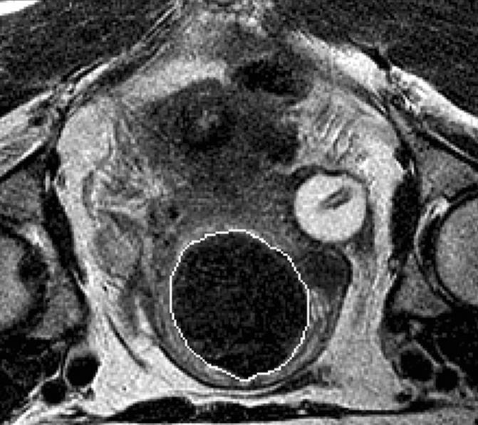
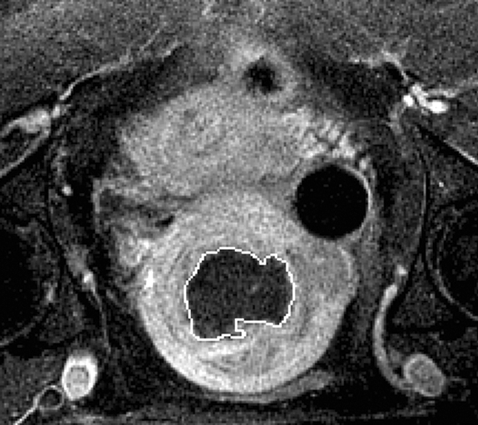
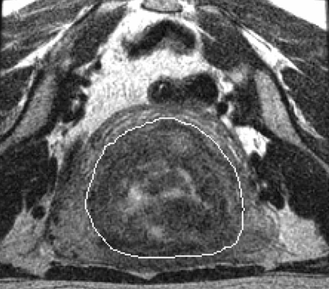
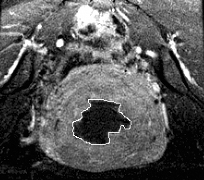
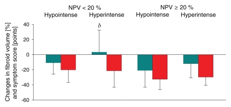
Materials and methods: This single-center retrospective analysis of 71 symptomatic fibroids in 66 women was approved by the institutional review board and was HIPAA-compliant. Patients were treated with MR imaging-guided focused ultrasound surgery. The volume of treated fibroid and nonperfused volume (NPV) were calculated with software, while symptom outcome was assessed with a symptom severity score (SSS). Fibroids were classified as hyperintense or hypointense relative to skeletal muscle on pretreatment T2-weighted MR images.
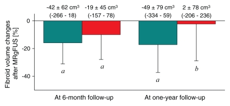
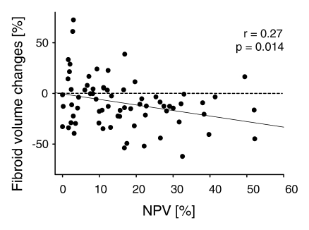
Results: Baseline volume of treated fibroids was 255.5 cm(3) +/- 201.7 (standard deviation), and baseline SSS was 61.5 +/- 14.9. Both pretreatment fibroid signal intensity (SI) and posttreatment NPV predicted 12-month volume reduction independently: Fibroids with an NPV of at least 20% or with low SI both showed significantly larger volume reduction (17.0% +/- 13.0 and 17.2% +/- 20.1, respectively) than fibroids with an NPV less than 20% or with high SI (10.7% +/- 18.2 and no significant change, respectively). Patients whose fibroids demonstrated an NPV of at least 20% also experienced a larger decrease in SSS than did patients with fibroids with an NPV less than 20% (50.1% +/- 19.8 vs 32.6% +/- 29.9).
Conclusion: Fibroids with low SI on pretreatment T2-weighted MR images were more likely to shrink than were ones with high SI. The larger the NPV immediately after treatment, the greater the volume reduction and symptom relief were. These findings may help both in selecting appropriate patients for MR-guided focused ultrasound surgery and in predicting patient outcome.
Figures
Similar articles
-
What predicts durable symptom relief of uterine fibroids treated with MRI-guided focused ultrasound? A multicenter trial in 8 academic centers.Eur Radiol. 2023 Nov;33(11):7360-7370. doi: 10.1007/s00330-023-09984-4. Epub 2023 Aug 9. Eur Radiol. 2023. PMID: 37553488 Clinical Trial.
-
Perfusion volume correlates, percentage of involution, and clinical efficacy at diverse follow-up survey times after MR-guided focused ultrasound surgery in uterine fibroids: first report in a Mexican mestizo population.Eur Radiol. 2015 Oct;25(10):2905-12. doi: 10.1007/s00330-015-3707-2. Epub 2015 Mar 26. Eur Radiol. 2015. PMID: 25809744
-
Feasibility of ultrasound-guided high intensity focused ultrasound ablating uterine fibroids with hyperintense on T2-weighted MR imaging.Eur J Radiol. 2013 Jan;82(1):e43-9. doi: 10.1016/j.ejrad.2012.08.020. Epub 2012 Sep 20. Eur J Radiol. 2013. PMID: 23000188
-
Volumetric MR-guided high-intensity focused ultrasound ablation with a one-layer strategy to treat large uterine fibroids: initial clinical outcomes.Radiology. 2012 May;263(2):600-9. doi: 10.1148/radiol.12111707. Epub 2012 Mar 8. Radiology. 2012. PMID: 22403170
-
Magnetic resonance-high intensity focused ultrasound (MR-HIFU) therapy of symptomatic uterine fibroids with unrestrictive treatment protocols: A systematic review and meta-analysis.Eur J Radiol. 2019 Nov;120:108700. doi: 10.1016/j.ejrad.2019.108700. Epub 2019 Oct 15. Eur J Radiol. 2019. PMID: 31634683
Cited by
-
Magnetic resonance temperature imaging-based quantification of blood flow-related energy losses.NMR Biomed. 2015 Jul;28(7):840-51. doi: 10.1002/nbm.3318. Epub 2015 May 14. NMR Biomed. 2015. PMID: 25973583 Free PMC article.
-
Assessment of acute thermal damage volumes in muscle using magnetization-prepared 3D T2 -weighted imaging following MRI-guided high-intensity focused ultrasound therapy.J Magn Reson Imaging. 2017 Aug;46(2):354-364. doi: 10.1002/jmri.25605. Epub 2017 Jan 9. J Magn Reson Imaging. 2017. PMID: 28067975 Free PMC article.
-
Preservation of the endometrial enhancement after magnetic resonance imaging-guided high-intensity focused ultrasound ablation of submucosal uterine fibroids.Eur Radiol. 2017 Sep;27(9):3956-3965. doi: 10.1007/s00330-017-4765-4. Epub 2017 Feb 16. Eur Radiol. 2017. PMID: 28210800
-
Magnetic Resonance-Guided High-Intensity Focused Ultrasound (MRgHIFU) Treatment of Symptomatic Uterine Fibroids: An Evidence-Based Analysis.Ont Health Technol Assess Ser. 2015 Mar 1;15(4):1-86. eCollection 2015. Ont Health Technol Assess Ser. 2015. PMID: 26357530 Free PMC article. Review.
-
Safety and Efficacy of High-intensity Focused Ultrasound Ablation for Patients with Submucosal Fibroids without Fertility Needs: A Single-center Real-world Data Retrospective Study.Gynecol Minim Invasive Ther. 2025 May 22;14(2):137-144. doi: 10.4103/gmit.GMIT-D-24-00045. eCollection 2025 Apr-Jun. Gynecol Minim Invasive Ther. 2025. PMID: 40521576 Free PMC article.
References
-
- Tempany CM, Stewart EA, McDannold N, Quade BJ, Jolesz FA, Hynynen K. MR imaging–guided focused ultrasound surgery of uterine leiomyomas: a feasibility study. Radiology 2003;226(3):897–905. - PubMed
-
- Stewart EA, Gedroyc WMW, Tempany CM, et al. Focused ultrasound treatment of uterine fibroid tumors: safety and feasibility of a noninvasive thermoablative technique. Am J Obstet Gynecol 2003;189(1):48–54. - PubMed
-
- Hindley J, Gedroyc WM, Regan L, et al. MRI-guidance of focused ultrasound therapy of uterine fibroids: early results. AJR Am J Roentgenol 2004;183(6):1713–1719. - PubMed
-
- Stewart EA, Rabinovici J, Tempany CM, et al. Clinical outcomes of focused ultrasound surgery for the treatment of uterine fibroids. Fertil Steril 2006;85(1):22–29. - PubMed
-
- Fennessy FM, Tempany CM, McDannold NJ, et al. Uterine leiomyomas: MR imaging–guided focused ultrasound surgery—results of different treatment protocols. Radiology 2007;243(3):885–893. - PubMed
Publication types
MeSH terms
Grants and funding
LinkOut - more resources
Full Text Sources
Medical
