

Targeted microbubbles for imaging tumor angiogenesis: assessment of whole-body biodistribution with dynamic micro-PET in mice
- PMID: 18695212
- PMCID: PMC2657857
- DOI: 10.1148/radiol.2491072050
Targeted microbubbles for imaging tumor angiogenesis: assessment of whole-body biodistribution with dynamic micro-PET in mice
Abstract
Purpose: To evaluate in vivo whole-body biodistribution of microbubbles (MBs) targeted to tumor angiogenesis-related vascular endothelial growth factor (VEGF) receptor 2 (VEGFR2) by using dynamic micro-positron emission tomography (PET) in living mice.
Materials and methods: Animal protocols were approved by the Institutional Administrative Panel on Laboratory Animal Care. Lipid-shell perfluorocarbon-filled MBs, targeted to VEGFR2 via anti-VEGFR2 antibodies, were radiolabeled by conjugating the radiofluorination agent N-succinimidyl-4-[(18)F]fluorobenzoate (SFB) to the anti-VEGFR2 antibodies. These MBs were then injected intravenously into nude mice (n = 4) bearing angiosarcomas, and the whole-body biodistribution of these probes was assessed for 60 minutes by using dynamic micro-PET. Results were compared with ex vivo gamma counting (n = 6) and immunofluorescence staining (n = 6). Control studies in angiosarcoma-bearing mice were performed with injection of the radiolabeled antibodies alone (n = 3) or free SFB (n = 3). A mixed-effects regression of MB accumulation on fixed effects of time and tissue type (tumor or muscle) and random effect of animal was performed.
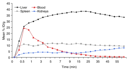
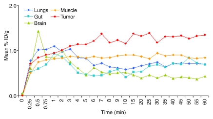
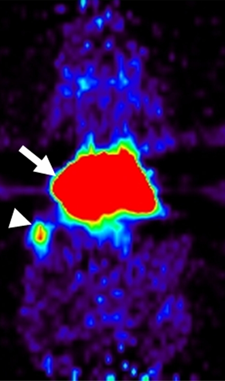
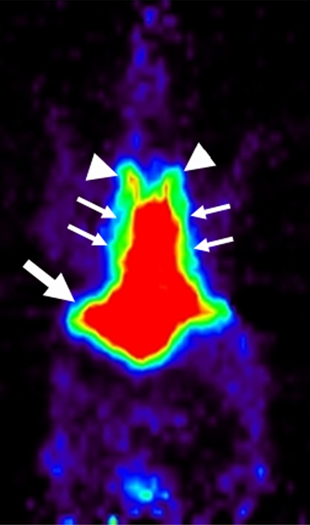
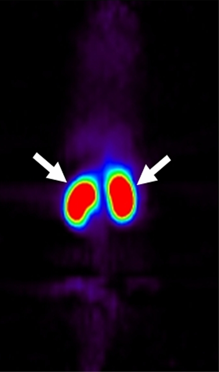
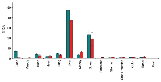
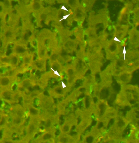
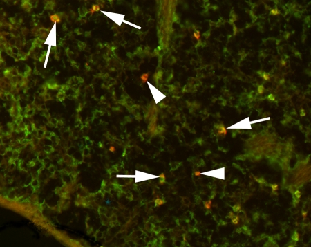
Results: VEGFR2-targeted MBs rapidly cleared from the blood circulation (50% blood clearance after approximately 3.5 minutes) and accumulated in the liver (mean, 33.4% injected dose [ID]/g +/- 13.7 [standard deviation] at 60 minutes) and spleen (mean, 9.3% ID/g +/- 6.5 at 60 minutes) on the basis of micro-PET imaging. These findings were confirmed with ex vivo gamma counting. Uptake of targeted MBs was significantly higher (P < .0001) in tumor than in adjacent skeletal muscle tissue. Immunofluorescence staining demonstrated accumulation of the targeted MBs within hepatic Kupffer cells and splenic macrophages. Biodistribution of the radiolabeled antibodies and free SFB differed from the distribution of the targeted MBs.
Conclusion: Dynamic micro-PET allows assessment of in vivo biodistribution of VEGFR2-targeted MBs.
(c) RSNA, 2008.
Figures
References
-
- Lindner JR. Microbubbles in medical imaging: current applications and future directions. Nat Rev Drug Discov 2004;3:527–532. - PubMed
-
- Willmann JK, van Bruggen N, Dinkelborg LM, Gambhir SS. Molecular imaging in drug development. Nat Rev Drug Discov 2008;7:591–607. - PubMed
-
- Ellegala DB, Leong-Poi H, Carpenter JE, et al. Imaging tumor angiogenesis with contrast ultrasound and microbubbles targeted to alpha(v)beta3. Circulation 2003;108:336–341. - PubMed
-
- Korpanty G, Carbon JG, Grayburn PA, Fleming JB, Brekken RA. Monitoring response to anticancer therapy by targeting microbubbles to tumor vasculature. Clin Cancer Res 2007;13:323–330. - PubMed
-
- Weller GE, Wong MK, Modzelewski RA, et al. Ultrasonic imaging of tumor angiogenesis using contrast microbubbles targeted via the tumor-binding peptide arginine-arginine-leucine. Cancer Res 2005;65:533–539. - PubMed
Publication types
MeSH terms
Substances
Grants and funding
LinkOut - more resources
Full Text Sources
Other Literature Sources
Miscellaneous
