. 2008 Aug 19;105(33):11691-6.
doi: 10.1073/pnas.0712239105. Epub 2008 Aug 11.

The identification of an osteoclastogenesis inhibitor through the inhibition of glyoxalase I

Affiliations

The identification of an osteoclastogenesis inhibitor through the inhibition of glyoxalase I

Makoto Kawatani et al. Proc Natl Acad Sci U S A. .

Abstract

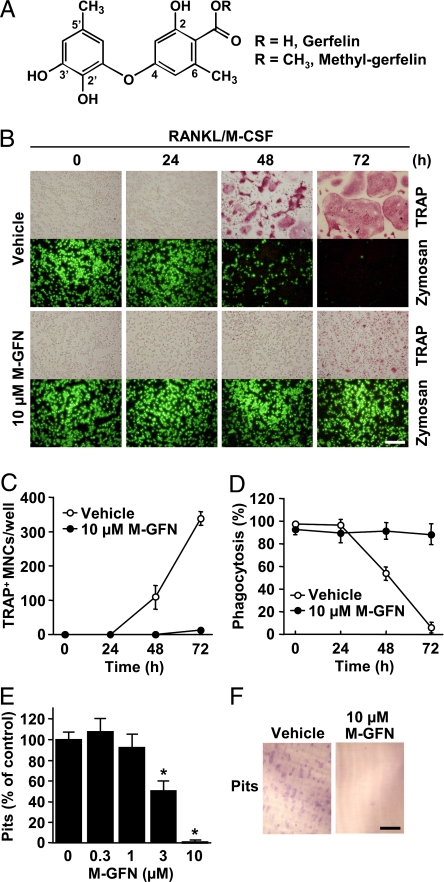
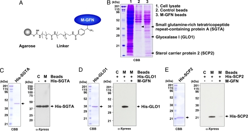
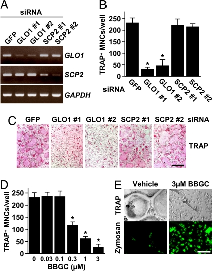
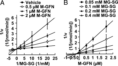
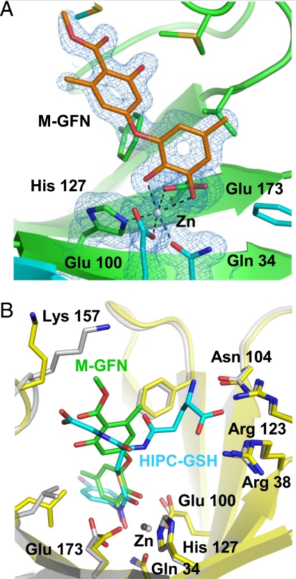
Osteoclasts, bone-resorptive multinucleated cells derived from hematopoietic stem cells, are associated with many bone-related diseases, such as osteoporosis. Osteoclast-targeting small-molecule inhibitors are valuable tools for studying osteoclast biology and for developing antiresorptive agents. Here, we have discovered that methyl-gerfelin (M-GFN), the methyl ester of the natural product gerfelin, suppresses osteoclastogenesis. By using M-GFN-immobilized beads, glyoxalase I (GLO1) was identified as an M-GFN-binding protein. GLO1 knockdown and treatment with an established GLO1 inhibitor in osteoclast progenitor cells interfered with osteoclast generation, suggesting that GLO1 activity is required for osteoclastogenesis. In cells, GLO1 plays a critical role in the detoxification of 2-oxoaldehydes, such as methylglyoxal. M-GFN inhibited the enzymatic activity of GLO1 in vitro and in situ. Furthermore, the cocrystal structure of the GLO1/M-GFN complex revealed the binding mode of M-GFN at the active site of GLO1. These results suggest that M-GFN targets GLO1, resulting in the inhibition of osteoclastogenesis.


Conflict of interest statement

The authors declare no conflict of interest.

Figures

Fig. 1.
Fig. 1.
M-GFN suppresses osteoclastogenesis with no influence on the phagocytic ability of BMMs. (A) Chemical structures of GFN and M-GFN. (B–D) BMMs were treated with M-GFN (10 μM) in the presence of RANKL and M-CSF for the indicated times. Then, cells were treated with fluorescein-conjugated zymosan A (FL-zymosan A), which is a model substrate to detect phagocytosis, and further stained for TRAP (B). TRAP+ multinucleated cells (MNCs) (C) and FL-zymosan A-incorporated phagocytic cells (D) were counted. Phagocytosis (%) means the ratio of the number of FL-zymosan A-incorporated cells to the total cell count. (E and F) Effect of M-GFN on the pit-forming activity of osteoclasts. BMMs were cultured on dentine slices with the indicated concentrations of M-GFN in the presence of RANKL and M-CSF for 72 h. Resorption pits on the slices, which are only formed by osteoclasts but not by BMMs, were stained with toluidine blue O (F), and the pits were counted (E). Data are shown as the mean ± SD (n = 4). *, P < 0.01 vs. Vehicle. (Scale bars, 200 μm.)
Fig. 2.
Fig. 2.
Identification of M-GFN-binding proteins. (A) Model structure of M-GFN beads. (B) Detection of the coprecipitated proteins for M-GFN beads from RAW264 cell lysates. RAW264 cell lysates were precleared with control beads and incubated with M-GFN beads. The reacted beads were washed, and the eluted proteins were subjected to SDS/PAGE and visualized by CBB staining. The coprecipitated proteins for M-GFN beads were identified as described in Materials and Methods. (C–E) Purified His-tagged SGTA (C), GLO1 (D), or SCP2 (E) protein was incubated with control beads and M-GFN beads in the presence or absence of M-GFN as a competitor. The reacted beads were washed, and the eluted proteins were immunoblotted with anti-Xpress Ab, which allows detection of recombinant His-tagged proteins containing the N-terminal leader peptide (Xpress epitope). C, Control beads; M, M-GFN beads.
Fig. 3.
Fig. 3.
GLO1 activity is required for osteoclastogenesis. (A–C) The knockdown of GLO1, but not of SCP2, fails to generate osteoclasts. (A) BMMs were transfected with siRNAs derived from mouse GLO1 and SCP2, or GFP, and then cultured in the presence of RANKL and M-CSF for 48 h. To confirm the depletion of these mRNAs, RT-PCR was performed. (B and C) BMMs were transfected with siRNAs derived from mouse GLO1 and SCP2, or GFP, and then cultured in the presence of RANKL and M-CSF for 72 h. Cells were fixed and stained for TRAP (C), and TRAP+ MNCs were counted (B). Data are shown as the mean ± SD (n = 5). *, P < 0.01 vs. GFP siRNA. (Scale bar, 500 μm.) (D and E) A known GLO1 inhibitor suppresses osteoclastogenesis. BMMs were treated with the indicated concentrations of BBGC in the presence of RANKL and M-CSF for 72 h. Then, cells were treated with FL-zymosan A and further stained for TRAP (E). TRAP+ MNCs were counted (D). Data are shown as the mean ± SD (n = 4). *, P < 0.01 vs. vehicle. (Scale bar, 150 μm.)
Fig. 4.
Fig. 4.
M-GFN inhibits GLO1 activity in a competitive manner. (A and B) Kinetic analysis of M-GFN against mouse His-GLO1. (A) Lineweaver–Burk plot of initial velocity vs. varying MG-SG concentrations. (B) Dixon plot of initial velocity vs. varying M-GFN concentrations. Data are shown as the mean ± SD (n = 3).
Fig. 5.
Fig. 5.
Cocrystal structure of GLO1 complexed with M-GFN. (A) Structure of M-GFN binding to the active site zinc ion of mouse GLO1. 2FobsFcalc electron density map contored at 1.0 sigma and overlaid on its structural model. Carbon atoms in one molecule of GLO1 dimer, the other molecule, and M-GFN are drawn in green, cyan, and orange, respectively. Gray sphere represents zinc ion. (B) Comparison of M-GFN/GLO1 complex and superimposed HIPC-GSH/GLO1 complex. M-GFN and HIPC-GSH molecules are shown as stick models in green and cyan, respectively. Carbon atoms of the protein (stick and ribbon model) and zinc ion in M-GFN/GLO1 model are drawn in yellow and dark gray, respectively, and those of HIPC-GSH/GLO1 are in gray. Oxygen, nitrogen, and sulfur atoms are drawn in red, blue, and gold, respectively, in both models. The half side of M-GFN partially overlaps the glycyl moiety of superimposed HIPC-GSH.

Similar articles

Cited by

References

    1. Boyle WJ, Simonet WS, Lacey DL. Osteoclast differentiation and activation. Nature. 2003;423:337–342. - PubMed
    1. Suda T, et al. Modulation of osteoclast differentiation and function by the new members of the tumor necrosis factor receptor and ligand families. Endocr Rev. 1999;20:345–357. - PubMed
    1. Hofbauer LC, Neubauer A, Heufelder AE. Receptor activator of nuclear factor-κB ligand and osteoprotegerin: Potential implications for the pathogenesis and treatment of malignant bone diseases. Cancer. 2001;92:460–470. - PubMed
    1. Rogers MJ. New insights into the molecular mechanisms of action of bisphosphonates. Curr Pharmacol Des. 2003;9:2643–2658. - PubMed
    1. Orcel P, Denne MA, de Vernejoul MC. Cyclosporin-A in vitro decreases bone resorption, osteoclast formation, and the fusion of cells of the monocyte-macrophage lineage. Endocrinology. 1991;128:1638–1646. - PubMed

Publication types

MeSH terms

Associated data