. 2008 Aug 19;105(33):11720-4.
doi: 10.1073/pnas.0804015105. Epub 2008 Aug 11.

Identification of a vesicular aspartate transporter

Affiliations

Identification of a vesicular aspartate transporter

Takaaki Miyaji et al. Proc Natl Acad Sci U S A. .

Abstract

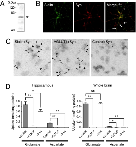
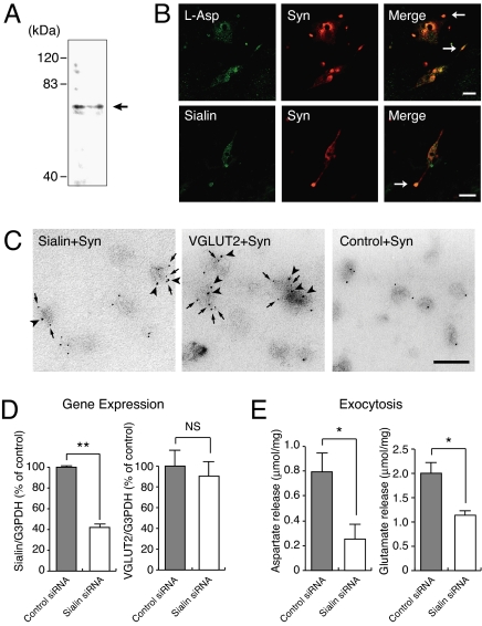
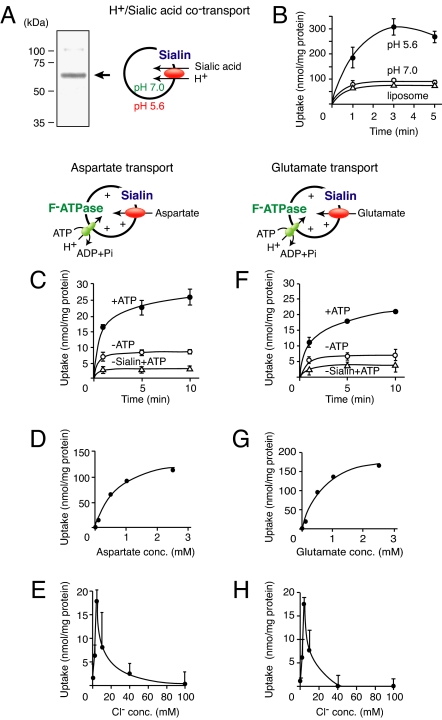
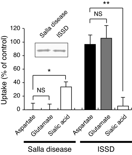
Aspartate is an excitatory amino acid that is costored with glutamate in synaptic vesicles of hippocampal neurons and synaptic-like microvesicles (SLMVs) of pinealocytes and is exocytosed and stimulates neighboring cells by binding to specific cell receptors. Although evidence increasingly supports the occurrence of aspartergic neurotransmission, this process is still debated because the mechanism for the vesicular storage of aspartate is unknown. Here, we show that sialin, a lysosomal H(+)/sialic acid cotransporter, is present in hippocampal synaptic vesicles and pineal SLMVs. RNA interference of sialin expression decreased exocytosis of aspartate and glutamate in pinealocytes. Proteoliposomes containing purified sialin actively accumulated aspartate and glutamate to a similar extent when inside positive membrane potential is imposed as the driving force. Sialin carrying a mutation found in people suffering from Salla disease (R39C) was completely devoid of aspartate and glutamate transport activity, although it retained appreciable H(+)/sialic acid cotransport activity. These results strongly suggest that sialin possesses dual physiological functions and acts as a vesicular aspartate/glutamate transporter. It is possible that people with Salla disease lose aspartergic (and also the associated glutamatergic) neurotransmission, and this could provide an explanation for why Salla disease causes severe neurological defects.


Conflict of interest statement

The authors declare no conflict of interest.

Figures

Fig. 1.
Fig. 1.
Phylogenetic tree of the SLC17 anion transporter family.
Fig. 2.
Fig. 2.
ATP-dependent uptake of aspartate was detected in sialin-containing synaptic vesicle preparation from hippocampus. (A) Immunoblotting revealed the presence of sialin in the hippocampal P2 fraction. The position of sialin is marked by an arrow. (B) Sialin is partially colocalized with synaptophysin. Cultured hippocampal neurons were double immunostained with antibodies against sialin and synaptophysin. (Scale bar, 10 μm.) (C) Double-labeling immunoelectron microscopy demonstrated the association of sialin with synaptophysin-containing vesicles. Samples were treated with sets of anti-synaptophysin monoclonal antibodies (5-nm particles, arrows) and anti-sialin serum (10-nm particles, arrowheads), anti-synaptophysin monoclonal antibodies (5-nm particles, arrows) and anti-VGLUT1 serum (10-nm particles, arrowheads), or anti-synaptophysin monoclonal antibodies (5-nm particles) and control serum (10-nm particles). (Scale bar, 100 nm.) (D) ATP-dependent uptake of aspartate and glutamate at 5 min by P2 fractions isolated from hippocampus and whole brain. Concentrations of CCCP and tHA were 1 μM and 5 mM, respectively.
Fig. 3.
Fig. 3.
Sialin was associated with pineal SLMVs and is involved in exocytosis of aspartate and glutamate. (A) Sialin was present in pineal SLMV fractions as revealed by immunoblotting. The position of sialin is marked by an arrow. (B) Sialin is colocalized with aspartate and synaptophysin. Cultured pinealocytes were double immunostained with antibodies against aspartate and synaptophysin (Upper) or sialin and synaptophysin (Lower). (Scale bar, 10 μm.) (C) Double-labeling immunoelectron microscopy demonstrated the association of sialin with synaptophysin. Samples were treated with sets of anti-synaptophysin monoclonal antibodies (5-nm particles, arrows) and anti-sialin serum (10-nm particles, arrowheads) or anti-synaptophysin monoclonal antibodies (5-nm particles, arrows) and anti-VGLUT2 serum (10-nm particles, arrowheads) or anti-synaptophysin monoclonal antibodies (5-nm particles) and control serum (10-nm particles). (Scale bar, 100 nm.) (D) RNAi directed against rat sialin decreased sialin expression without affecting the expression of VGLUT2. Quantitative analysis for sialin and VGLUT2 mRNA levels was performed by real-time PCR. (E) Exocytosis of aspartate and glutamate after 20-min incubation upon KCl stimulation from control siRNA-treated pinealocytes and sialin siRNA-treated pinealocytes.
Fig. 4.
Fig. 4.
Sialin is a vesicular aspartate/glutamate transporter. (A) Purified mouse sialin (10 μg of protein) was visualized by Coomassie brilliant blue staining after SDS/PAGE. The position of sialin is indicated by an arrow. (B) H+/sialic acid cotransport by the proteoliposomes was assayed by the pH jump method (acidic outside). (C–E) Sialin is a vesicular aspartate transporter. (C) Time course of ATP-dependent aspartate uptake by proteoliposomes containing reconstituted mouse sialin and bacterial F-ATPase. The reaction was started by adding radiolabeled aspartate to a final concentration of 100 μM in the presence (filled circles) or absence (open circles) of 2 mM ATP. (D) Dose dependence of aspartate uptake at 1 min. (E) Chloride ion dependence of aspartate uptake at 5 min was measured in buffer containing KCl at the indicated concentrations. (F–H) Sialin is a VGLUT. Radiolabeled glutamate was used instead of radiolabeled aspartate. (F) Time course. (G) Dose dependence. (H) Chloride ion dependence.
Fig. 5.
Fig. 5.
Transport activity of mutant sialins that are associated with Salla disease (R39C) and ISSD (H183R). (Inset) Coomassie brilliant blue-stained mutant protein (10 μg). Δψ-dependent uptake of aspartate and glutamate at 5 min and H+/sialic acid cotransport by proteoliposomes at 3 min is shown. Control activity corresponded to that of wild-type sialin (Fig. 4).

References

    1. Maycox PR, Hell JW, Jahn R. Amino acid neurotransmission: Spotlight on synaptic vesicles. Trends Neurosci. 1990;13:83–87. - PubMed
    1. Schuldiner S, Shirvan A, Linial M. Vesicular neurotransmitter transporters: From bacteria to humans. Physiol Rev. 1995;75:369–392. - PubMed
    1. Gasnier B. The SLC32 transporter, a key protein for the synaptic release of inhibitory amino acids. Pflügers Arch. 2004;447:756–759. - PubMed
    1. Reimer RJ, Edwards RH. Organic anion transport is the primary function of the SLC17/type I phosphate transporter family. Pflügers Arch. 2004;447:629–635. - PubMed
    1. Bliss TVP, Collingridge GL. A synaptic model of memory: Long-term potentiation in the hippocampus. Nature. 1993;361:31–39. - PubMed

Publication types

MeSH terms