. 2008 Oct 20;510(6):561-80.
doi: 10.1002/cne.21801.

Minimal latency to hippocampal epileptogenesis and clinical epilepsy after perforant pathway stimulation-induced status epilepticus in awake rats

Affiliations

Minimal latency to hippocampal epileptogenesis and clinical epilepsy after perforant pathway stimulation-induced status epilepticus in awake rats

Argyle V Bumanglag et al. J Comp Neurol. .

Abstract

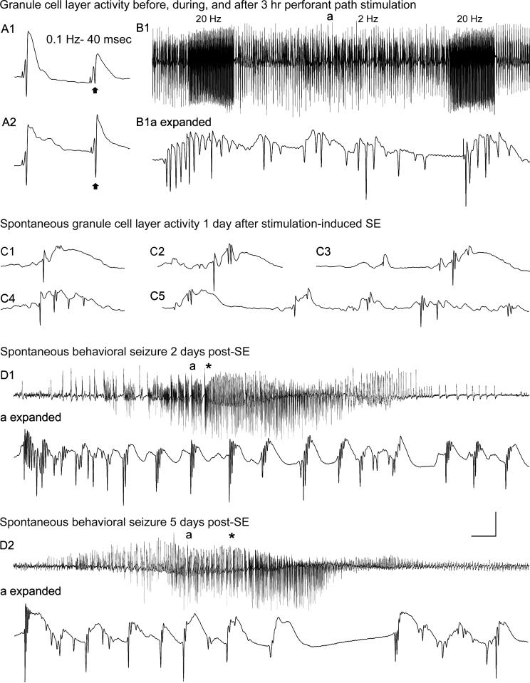
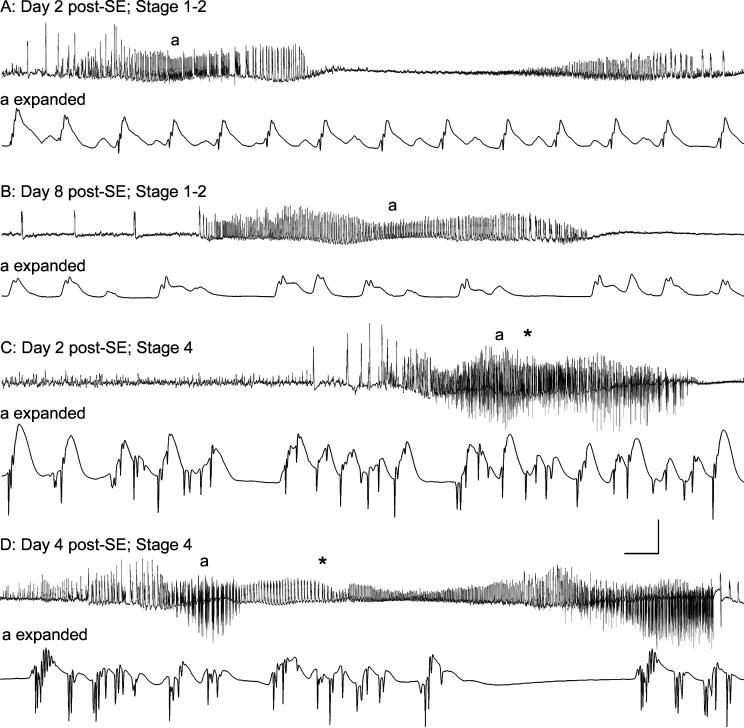
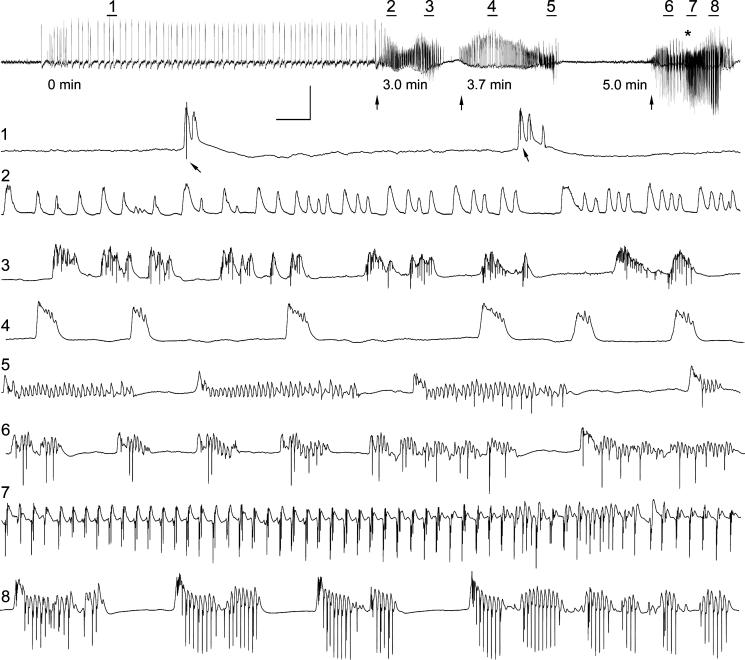
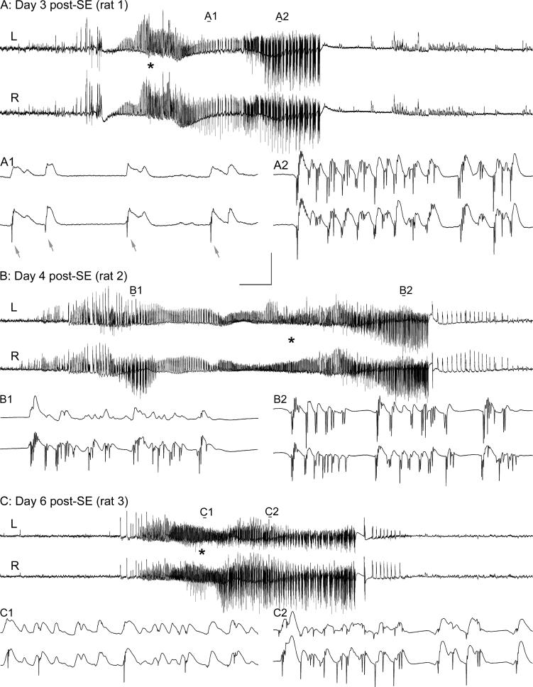
Hippocampal epileptogenesis is hypothesized to involve secondary mechanisms triggered by initial brain injury. Chemoconvulsant-induced status epilepticus has been used to identify secondary epileptogenic mechanisms under the assumption that a seizure-free, preepileptic "latent period" exists that is long enough to accommodate delayed mechanisms. The latent period is difficult to assess experimentally because early spontaneous seizures may be caused or influenced by residual chemoconvulsant that masks the true duration of the epileptogenic process. To avoid the use of chemoconvulsants and determine the latency to hippocampal epileptogenesis and clinical epilepsy, we developed an electrical stimulation-based method to evoke hippocampal discharges in awake rats and produce hippocampal injury and hippocampal-onset epilepsy reliably. Continuous video monitoring and granule cell layer recording determined whether hippocampal epileptogenesis develops immediately or long after injury. Bilateral perforant pathway stimulation for 3 hours evoked granule cell epileptiform discharges and convulsive status epilepticus with minimal lethality. Spontaneous stage 3-5 behavioral seizures reliably developed within 3 days poststimulation, and all 72 spontaneous behavioral seizures recorded in 10 animals were preceded by spontaneous granule cell epileptiform discharges. Histological analysis confirmed a reproducible pattern of limited hippocampal and extrahippocampal injury, including an extensive bilateral loss of hilar neurons throughout the hippocampal longitudinal axis. These results indicate that hippocampal epileptogenesis after convulsive status epilepticus is an immediate network defect coincident with neuron loss or other early changes. We hypothesize that the latent period is directly related and inversely proportional to the extent of neuron loss in brain regions involved in seizure initiation, spread, and clinical expression.


Figures

Figure 1
Figure 1
Dentate granule cell excitability and spontaneous activity before, and 1−5 days after, 3 hr of perforant pathway stimulation-induced convulsive status epilepticus (SE). (A1) Before SE, paired-pulse perforant pathway stimulation at 0.1 Hz and an inter-stimulus interval of 40 msec evokes granule cell responses that exhibit partial suppression of the amplitude of the second population spike (arrow). (A2) Three days after 3 hr of SE, the identical afferent stimulation failed to suppress the second population spike (arrow). (B1) Granule cell layer activity during 3 hr of perforant pathway stimulation in the same awake rat. The stimulation paradigm involved continuous stimulation at 2 Hz with paired pulses delivered at a 40 msec interpulse interval, plus 10 sec-long 20 Hz trains delivered once per minute. Note the morphology of the granule cell epileptiform discharges during the 2 Hz inter-train interval (a) in the expanded trace (B1a expanded). (C) On the first day after 3 hr of stimulation-induced SE, a granule cell layer electrode recorded spontaneous granule cell field “EPSPs” and population spikes that closely resemble the evoked responses in (A). (D) Granule cell layer activity during spontaneous behavioral seizures during the first week post-SE. (D1) On day 2 post-SE, granule cell layer activity amplitude increased before the behavioral onset of the second behavioral seizure on that day (marked by asterisk). (D1a expanded) expanded trace of the region above marked “a,” showing that the high-amplitude activity in (D1) consisted of granule cell epileptiform discharges that preceded the behavioral seizure-onset (asterisk). (D2) Three days later, the fourth spontaneous behavioral seizure exhibited nearly identical features including high-frequency granule cell epileptiform discharges (D2a expanded) that preceded the behavioral seizure-onset (asterisk). Calibration bars: (A) 14 msec and 9 mV; (B1) 7 sec and 9 mV; (B1 expanded) 46 msec and 9 mV; (C) 40 msec and 9 mV; (D1) 3.4 sec and 9 mV; (D1 expanded) 53 msec and 9 mV; (D2) 3.4 sec and 9 mV; (D2 expanded) 60 msec and 9 mV.
Figure 2
Figure 2
Correlation between spontaneous granule cell layer activity and behavioral seizure expression in an epileptic rat 2−8 days after 3 hr of perforant pathway stimulation-induced convulsive status epilepticus (SE). Two focal (subclinical) seizures (A) and (B) on days 2 and 8 post-SE, and two Stage 4 behavioral seizures (C) and (D) on days 2 and 4 post-SE. (A) On Day 2 post-SE, spontaneous high-amplitude activity, consisting of field “EPSPs” and small amplitude population spikes, was recorded from the granule cell layer electrode. During this spontaneous event, the animal exhibited only a frozen stare, followed by stereotyped chewing movements. (B) A similar spontaneous granule cell layer event was recorded on Day 8 post-SE, which was also associated with staring and stereotyped head movements only. (C) and (D) on Days 2 and 4 post-SE, spontaneous granule cell layer events included larger amplitude, downwardly deflected population spikes, and these events were invariably followed by Stage 3−5 behavioral seizures. Note that high-frequency spiking began prior to the first behavioral manifestation (asterisks in C and D) of each behavioral seizure. These events from a single rat (different from the rat shown in Fig. 1) are representative of all 72 Stage 3−5 behavioral seizures recorded in 10 chronically-implanted, continuously monitored rats. Calibration bars: (A) 1.4 sec and 9 mV; (A expanded) 56 msec and 9 mV; (B) 3.2 sec and 9 mV; (B expanded) 56 msec and 9 mV (C) 3.2 sec and 9 mV; (C expanded) 56 msec and 9 mV; (D) 4.5 sec and 9 mV; (D expanded) 56 msec and 9 mV.
Figure 3
Figure 3
Granule cell layer activity and clinical seizure expression during one 6-minute period in an awake epileptic rat 9 days after 3 hr of perforant pathway stimulation-induced convulsive status epilepticus (SE). Top trace: 6 min of granule cell layer activity showing 3 distinct events (arrows), the last of which culminated in a Stage 4 spontaneous epileptic seizure (forepaw clonus and rearing) that began (asterisk) ∼19 sec after the onset of high amplitude granule cell population spikes. Prior to the first onset of high frequency granule cell layer activity at the 3 min marker (first arrow), high amplitude, low frequency activity consisted of positive-going waves with occasional superimposed granule cell population spikes (arrows in expanded trace 1). During the first of 3 distinct, high-frequency granule cell layer events, positive-going “field EPSPs” and population spikes were recorded (traces 2 and 3, respectively). These granule cell layer discharges lacked large amplitude granule cell population spikes that extended below baseline, and these potentials were not associated with a behavioral seizure. A second similar event began 40 sec later, and contained mostly positive-going events (traces 4 and 5) that were not associated with a behavioral seizure. The third event consisted of large amplitude, high-frequency granule cell population spikes that extended far below baseline (traces 6−8), and these features were uniquely associated with the onset of a spontaneous Stage 4 epileptic seizure. Note that forepaw clonus (asterisk in top trace) began ∼15 sec after the onset of high amplitude granule cell population spikes shown in trace 6. The top trace is 6.0 min in duration, and expanded traces 1−8 are 4.4 sec in duration. Calibration bars: top trace, 16.8 sec and 12.5 mV; traces 1−8: 205 msec and 12.5 mV.
Figure 4
Figure 4
Bilateral synchrony of spontaneous granule cell layer epileptiform discharges in three different epileptic rats. Bilateral recording electrodes with their tips in the dentate granule cell layers recorded highly synchronous spontaneous activity in all animals subjected to 3 hr of perforant pathway stimulation-induced convulsive SE. (A): Prior to a Stage 3−5 behavioral seizure onset, bilateral recording electrodes recorded superficially identical granule cell layer activity (top traces; 2.0 min in duration). Expanded views (all are 0.9 sec in duration) of two segments of these spontaneous discharges reveal highly synchronized, but clearly not identical, discharges. Note population spikes on the lower traces (arrows in A1), but only field depolarizations in the simultaneously recorded top trace in (A1). During the high frequency granule cell layer discharges (A2), spiking was highly synchronous but not identical. (B) and (C): Similar events recorded in two other epileptic rats 4 and 6 days post-SE, respectively. Note that these highly synchronized discharges are representative of all 72 spontaneous seizures recorded. Calibration bars: unexpanded traces top trace, 7.3 sec and 12.5 mV; expanded traces: 114 msec and 12.5 mV.
Figure 5
Figure 5
Acute Fluoro-Jade B (FJB) staining showing neurodegeneration 4 days after 3 hr of perforant pathway stimulation-induced SE. (A) FJB fluorescence; (B and C) grayscale, inverted image of the same horizontal brain section. Note selective degeneration of neurons in the dentate hilar region (C-1), hippocampal area CA3a (C-2), entorhinal cortex layer III (C-3), perirhinal cortex (C-4 and 5), layer II throughout the neocortex (C-6), the parafascicular thalamic nucleus (C-7), the intermediodorsal-, mediodorsal-, paratenial-, paraventricular-, and centralmedial thalamic nuclei (C-8), lateral septum (C-9), lateral caudate/putamen (C-10), infralimbic cortex (C-11), and deep pyramidal cells of the agranular insular cortex (C-12). Calibration bar: 2mm in A and C; 1mm in B.
Figure 6
Figure 6
Acute neurodegeneration in hippocampus and entorhinal cortex 4 days after 3 hr of perforant pathway stimulation-induced SE. (A) Coronal section of a sham control rat (implanted, no SE) showing that the presence of the hippocampal recording electrode and the control treatment (electrode implantation, stimulation for 3 hr at 0.1 Hz, halothane anesthesia, and subanesthetic urethane treatment) produced no detectable hippocampal Fluoro-Jade B (FJB) staining (grayscale, inverted image as shown in preceding figure). (B) Coronal section of a stimulated rat (same rat as in the preceding figure) showing acute injury in the dorsal hippocampus 4 days after 3 hr of perforant pathway stimulation-induced SE. Note FJB-positive cells in the hilus, area CA3a and CA3c, and area CA1. (C) Horizontal brain section from the same rat stained with FJB, showing selective degeneration of neurons in the entorhinal cortex (wide arrow), the dentate hilus (h), and the dentate granule cell layer (gc). Note also the degenerating terminals in the inner dentate molecular layer (thin arrows) that originate from the degenerating hilar mossy cells. (D) Horizontal section from a sham control animal after immunostaining for the neuronal marker NeuN. (E) A horizontal section adjacent to the section shown in (C) after NeuN immunostaining showing the loss of NeuN immunoreactivity of neurons in the hilus (h) and entorhinal cortex. Calibration bar: 1mm in A and B; 400 μm in C-E.
Figure 7
Figure 7
Neuronal injury and loss in the dentate hilus and entorhinal cortex after 3 hr of perforant pathway stimulation-induced convulsive SE. Nissl-stained sections adjacent to those shown in the previous figure, in which NeuN immunostaining illustrated the apparent loss of hilar neurons. (A) Nissl-stained horizontal section from a sham control animal. (B) 4 days post-SE. Note the extensive loss of large Nissl-stained hilar neurons (h) and neurons in the entorhinal cortex (arrow). (C) and (D) Nissl-stained coronal sections from a sham control animal (C) and a stimulated animal 42 days post-SE (D). Note the extensive loss of hilar neurons (h). Calibration bar: 400μm in A and B; 285 μm in C and D.
Figure 8
Figure 8
Neuron loss 42 days after 3 hr of perforant pathway stimulation-induced SE. (A) Dorsal hippocampal dentate gyrus in a sham control section; NeuN immunostaining. (B) Dentate gyrus in a coronal section 42 days after 3 hr of perforant pathway stimulation-induced SE. Note extensive loss of NeuN-positive hilar neurons, but minimal apparent loss of CA3c pyramidal cells. (C) and (E) Low and higher magnification views of a horizontal section from the control brain. (D) and (F) Low and higher magnification views of a horizontal section 42 days post-SE, showing hilar neuron loss in the ventral dentate gyrus (open arrows) and the loss of entorhinal cortex neurons (thin arrows). Also note in (D) relatively subtle CA3 pyramidal neuron loss (wide arrow). Calibration bar: 400 μm in A and B; 2mm in C and D; 1mm in E and F.

Similar articles

Cited by

References

    1. Andersen P, Holmqvist B, Voorhoeve PE. Entorhinal activation of dentate granule cells. Acta Physiol Scand. 1966;66:448–460. - PubMed
    1. Arida RM, Scorza FA, Peres CA, Cavalheiro EA. The course of untreated seizures in the pilocarpine model of epilepsy. Epilepsy Res. 1999;34:99–107. - PubMed
    1. Bertram E. The relevance of kindling for human epilepsy. Epilepsia. 2007;48(Suppl 2):65–74. - PubMed
    1. Bertram EH, Cornett J. The ontogeny of seizures in a rat model of limbic epilepsy: evidence for a kindling process in the development of chronic spontaneous seizures. Brain Res. 1993;625:295–300. - PubMed
    1. Bertram EH, Cornett JF. The evolution of a rat model of chronic spontaneous limbic seizures. Brain Res. 1994;661:157–162. - PubMed

Publication types