Review
. 2008 Oct;237(10):2667-75.
doi: 10.1002/dvdy.21637.

Embryonic chick corneal epithelium: a model system for exploring cell-matrix interactions

Affiliations
Review

Embryonic chick corneal epithelium: a model system for exploring cell-matrix interactions

Kathy K H Svoboda et al. Dev Dyn. 2008 Oct.

Abstract

In her initial research, Elizabeth D. Hay studied amphibian limb regeneration, but later switched her focus, and for the remainder of her career addressed the role of the extracellular matrix (ECM) in regulating embryonic morphogenesis. Much of that work used the embryonic chick corneal epithelial model. This review highlights many of the discoveries that she made using this model. Hay was the first to show that embryonic corneal epithelial cells produce fibrillar collagen. Her lab was among the first to demonstrate that corneal epithelial cells respond to a collagenous substrate by increasing ECM production, and that purified ECM molecules, added to cultures of epithelial sheets, induce a reorganization of the actin cytoskeleton. These data led to the first theories of cell-matrix interactions, illustrated in a 'hands across the membrane' sketch drawn by Hay. Recent work with the epithelial sheet model system has elucidated many of the signal transduction pathways required for actin reorganization in response to the ECM. In all, this body of work has amply supported Hay's belief that the embryonic corneal epithelium is a powerful model system for exploring the role of the ECM in regulating the cytoskeleton, in directing cell migration, and in profoundly influencing cell growth and differentiation during development.


Figures

Fig. 1
Fig. 1
The culture system developed by Dodson, Meier, and Hay placing epithelium on killed lens capsule. A: Epithelium cultured directly on lens capsule. B: Epithelium separated from lens capsule by a Nucleopore filter barrier. Reprinted from Meier and Hay, 1975. C: Transmission electron micrograph of a 24-hr culture showing epithelial cell processes extending into the filter. Reprinted from Hay and Meier, 1976. D: Corneal epithelia grown directly on one Nucleopore filter, 0.8 μm pore size (bottom curve), on stacks of 0.8μm pore size filters with lens capsule beneath the filter, or directly on lens capsule (top curve). Cultures were grown in the presence of 5μCi/ml H3 proline and harvested at various times up to 24 hr. The bottom curve demonstrates the base line level of collagen synthesis, while the top curve corresponds to the “induced state.” Stimulation of synthetic activity decreased with the increasing filter thickness, consistent with the observation that the cell processes traversed the filter contacting the lens capsule. Vertical bars = standard deviation for four assays. Reprinted from Meier and Hay (1975).
Fig. 2
Fig. 2
Diagrams and electron micrographs illustrating the effects of different experimental conditions on the organization of the basal cell surface. A: The basal surface blebbed when the basal lamina was removed by EDTA or enzyme treatment, and the blebbing persisted on Millipore filters in the presence of nonmatrix proteins or glycosaminoglycans. B: Soluble collagens, fibronectin and laminin added to the medium stimulated the bleb retraction and reformation of the basal actin cytoskeleton. Reprinted from Sugure and Hay (1982). C: Corneal epithelial tissues were detergent extracted and treated with S-1 fragments of heavy meromyosin before EM preparation. C-1 is a typical bleb demonstrating a core of actin filaments decorated with myosin S-1 fragments, which aligns on the actin filaments indicating the polarity of the filament that is pointing toward the plasma membrane (indicated by small arrows) and some apparently inserting into a dense plaque (dp). Intermediate filaments (IF) were in the basal cell area. C-2 is a smaller bleb with some actin filaments parallel to the cell surface (arrowheads) and others perpendicular to the surface. C-3 presents a cell 6 hr after immersion in soluble type IV collagen (100 μg/ml). The actin bundle (MF) was in the basal compartment of this slightly tangential section containing a damaged plasma membrane (PM). The actin filaments were organized in opposite directions (small arrows). Scale bars = 0.2μm. Reprinted from Sugrue and Hay (1982). D. Hay's concept drawing illustrating the influence of the ECM on the actin cytoskeleton. It was used in lab meetings and titled “Hands across the membrane.”
Fig. 3
Fig. 3
Diagrams of several models depicting the possible relation of extracellular matrix (ECM) molecules to the cell surface. All models envision plasma membrane receptors for one or more molecules. CO, collagen; FN fibronectin; PG, proteoglycans; HA, hyaluronic acid. A: Model based on the data from Sugrue and Hay (1982). B: Model based on data from Singer (1979) and Kleinman et al. (1981). C: Model of FN interaction with fibroblasts by Hynes et al. (1982). D: Model of glycosaminoglycans and the mesenchymal cell surface from Toole (1981). Reprinted from Hay, (1981a,b).
Fig. 4
Fig. 4
Confocal microscopy of actin in corneal epithelial sheets. Schematic drawing of embryonic corneal epithelia isolation and culture. A: Epithelia were isolated as a sheet of tissue and placed on a polycarbonate filter, basal side down, then cultured at the air–media interface, B: Epithelia isolated without the basal lamina (−BL) extended basal cellular processes termed “blebs.” C: Sheets of corneal epithelia were stained with fluorescent phalloidin to label all filamentous actin (F-actin). In single confocal optical sections, the blebs are punctate spots in the plane of the basal cells. The cortical actin near cell membranes is also in the micrograph. D: If the tissue is cultured with soluble extracellular matrix (ECM) molecules, the basal actin reorganizes into an actin cortical mat (ACM) that phalloidin labels as bundles of F-actin that align from cell to cell after COL treatment for 2 hr. Scale bar = 10 μm. Reprinted from Svoboda et al. (1999b).
Fig. 5
Fig. 5
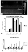
Confocal images of epithelia grown on filters with fluorescently labeled matrix components. A,B: Corneal epithelia were isolated without the basal lamina (−BL), incubated with media containing fluorescein isothiocyanate–fibronectin (FITC-FN) for short time intervals (15 or 30 min.). The epithelia were rinsed, fixed and viewed on the confocal microscope in the xz optical plane. The intensity of bound FITC-labeled FN was recorded at the basal cell surface in a nonuniform pattern. The intensity level of FITC-FN increased from 0 to 30 min as analyzed with NIH Image. C: Corneal epithelia −BL were isolated, incubated with media containing FN or COL for a short time interval (0, 10, 15, 20, or 30 min). The cultures were rinsed and immersed in control non-ECM media, and then incubated for a total time of 2 hr. Samples were fixed and stained with fluorescent phalloidin, and then groups coded (numbered) and scored by at least two people as +, +/−, or −actin cortical mat (ACM). The experiments were repeated three times, with an n = 5–7 epithelia/group. Folded and damaged tissue was not scored. Greater than 80% of the epithelia responded to the ECM molecules within 20-30 min, 22% with 10 min. D: Tritium labeled collagen was added to cultured epithelium in the presence of unlabeled collagen (1, 10, 50, 100 or 500 μg/ml) for 30, 60 or 120 min. Increased binding of 3H-collagen was observed up to 100 μg/ml but binding decreased at 500 μg/ml, suggesting competition at high levels of cold collagen. Reprinted from Svoboda et al. (1999b).

Similar articles

Cited by

References

    1. Ali IU, Hynes RO, Ali IU, Hynes RO. Effects of LETS glycoprotein on cell motility. Cell. 1978;14:439–446. - PubMed
    1. Bernfield MR. Collagen synthesis during epitheliomesenchymal interactions. Dev Biol. 1970;22:213–231. - PubMed
    1. Chu CL, Reenstra RW, Orlow DL, Svoboda KKH. Erk and PI3 kinase are necessary for collagen binding and actin reorganization in avian corneal epithelia. Invest Ophthalmol Vis Sci. 2000;41:3374–3382. - PMC - PubMed
    1. Coulombre AJ, Coulmbre JL. Corneal development. II. Transparency changes during rapid hydration. Am J Ophthalmol. 1958;46:276–280. discussion 281. - PubMed
    1. Dodson JW. The differentiation of epidermis. I. The interrelationship of epidermis and dermis on embryonic chicken skin. J Embryol Exp Morphol. 1967;17:83–105. - PubMed

Publication types

LinkOut - more resources