

A feasibility study to calculate unshielded fetal doses to pregnant patients in 6-MV photon treatments using Monte Carlo methods and anatomically realistic phantoms
- PMID: 18697528
- PMCID: PMC2809713
- DOI: 10.1118/1.2938519
A feasibility study to calculate unshielded fetal doses to pregnant patients in 6-MV photon treatments using Monte Carlo methods and anatomically realistic phantoms
Abstract
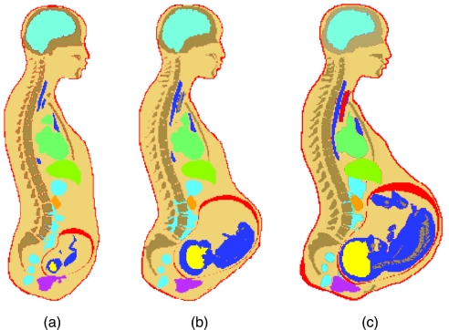
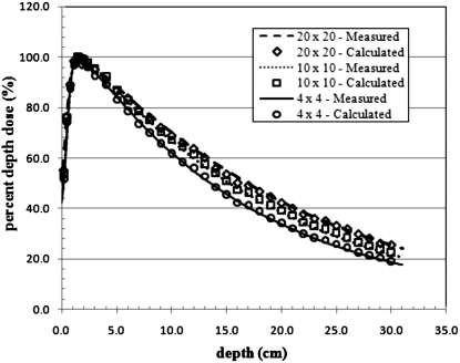
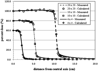
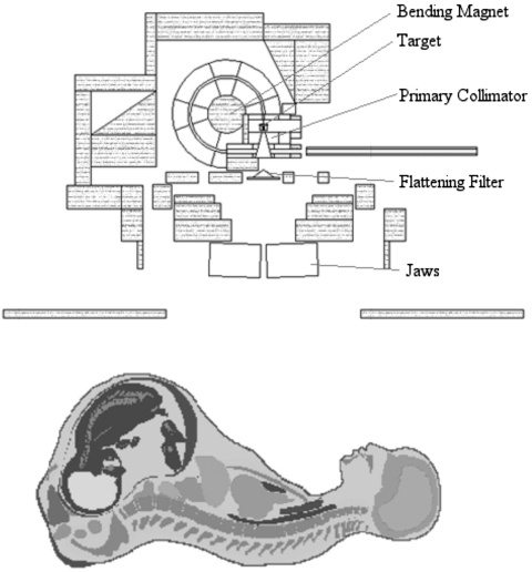
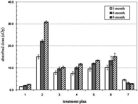
A Monte Carlo-based procedure to assess fetal doses from 6-MV external photon beam radiation treatments has been developed to improve upon existing techniques that are based on AAPM Task Group Report 36 published in 1995 [M. Stovall et al., Med. Phys. 22, 63-82 (1995)]. Anatomically realistic models of the pregnant patient representing 3-, 6-, and 9-month gestational stages were implemented into the MCNPX code together with a detailed accelerator model that is capable of simulating scattered and leakage radiation from the accelerator head. Absorbed doses to the fetus were calculated for six different treatment plans for sites above the fetus and one treatment plan for fibrosarcoma in the knee. For treatment plans above the fetus, the fetal doses tended to increase with increasing stage of gestation. This was due to the decrease in distance between the fetal body and field edge with increasing stage of gestation. For the treatment field below the fetus, the absorbed doses tended to decrease with increasing gestational stage of the pregnant patient, due to the increasing size of the fetus and relative constant distance between the field edge and fetal body for each stage. The absorbed doses to the fetus for all treatment plans ranged from a maximum of 30.9 cGy to the 9-month fetus to 1.53 cGy to the 3-month fetus. The study demonstrates the feasibility to accurately determine the absorbed organ doses in the mother and fetus as part of the treatment planning and eventually in risk management.
Figures

References
-
- Magne N., Marcie S., Pignol J., Casagrande F., and Lagrange J., “Radiotherapy for a solitary brain metastasis during pregnancy: A method for reducing fetal dose,” Br. J. Radiol. 74, 638–641 (2001). - PubMed
-
- Mazonakis M., Damilakis J., Theoharopoulos N., Varberis H., and Gourtsoyiannis N., “Brain radiotherapy during pregnancy: An analysis of conceptus dose using anthropomorphic phantoms,” Br. J. Radiol. 72, 274–278 (1999). - PubMed
Publication types
MeSH terms
Grants and funding
LinkOut - more resources
Full Text Sources
Medical
