. 2008 Sep;135(17):2905-16.
doi: 10.1242/dev.019778.

Dlx genes pattern mammalian jaw primordium by regulating both lower jaw-specific and upper jaw-specific genetic programs

Affiliations

Dlx genes pattern mammalian jaw primordium by regulating both lower jaw-specific and upper jaw-specific genetic programs

Juhee Jeong et al. Development. 2008 Sep.

Abstract

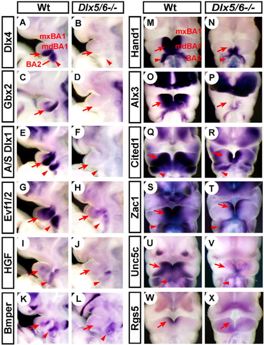
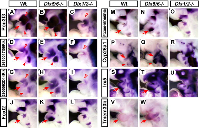
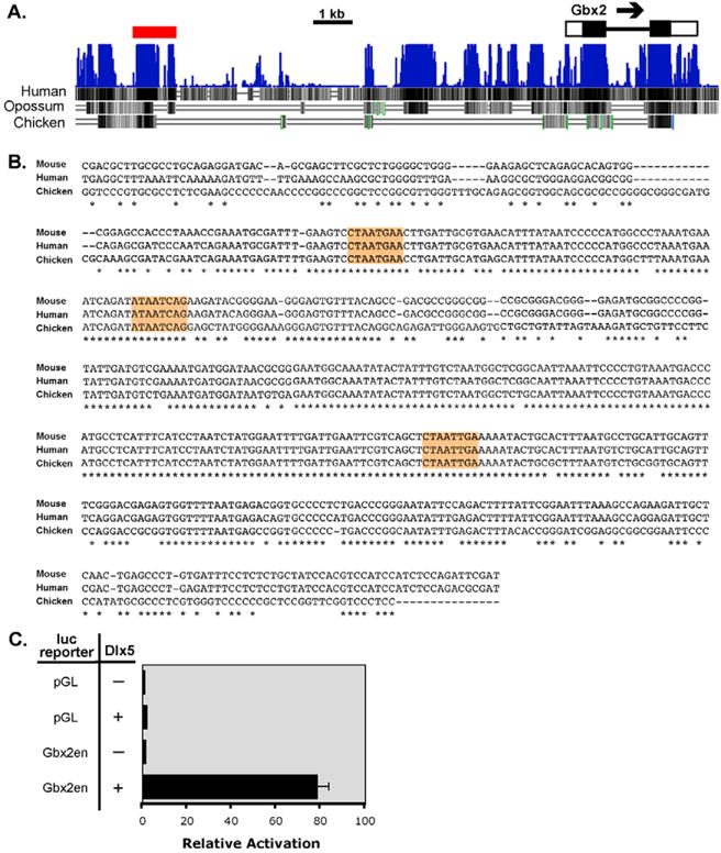
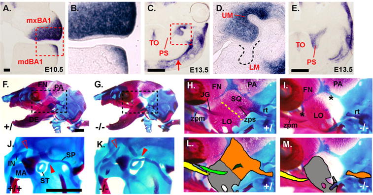
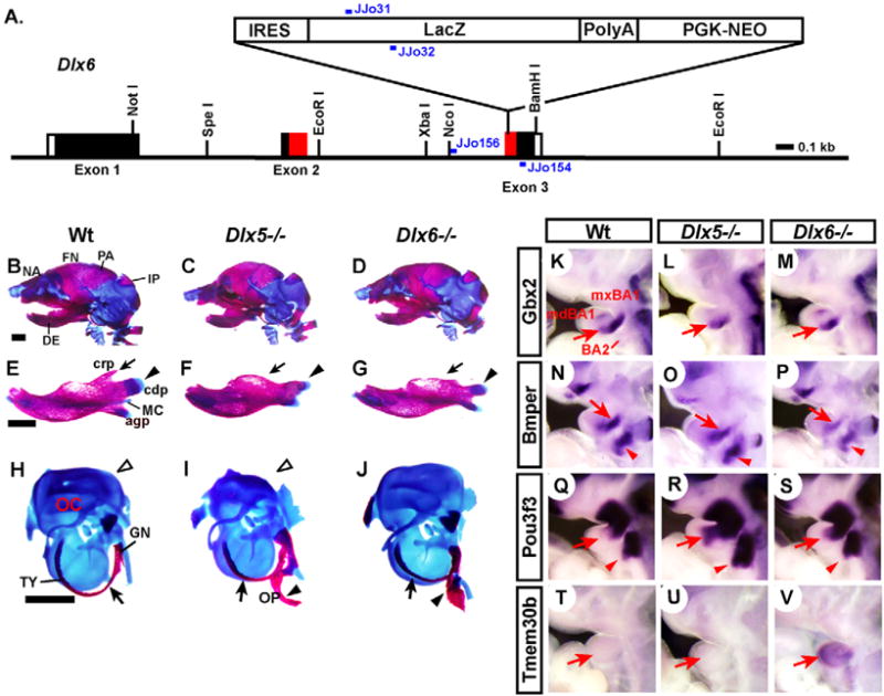
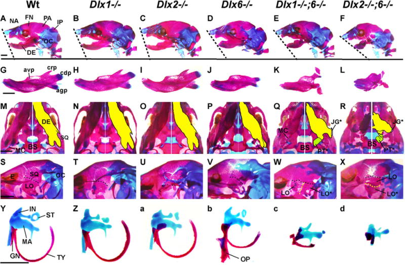
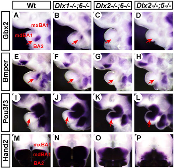
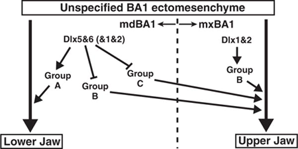
Dlx transcription factors are implicated in patterning the mammalian jaw, based on their nested expression patterns in the first branchial arch (primordium for jaw) and mutant phenotypes; inactivation of Dlx1 and Dlx2 (Dlx1/2-/-) causes defects in the upper jaw, whereas Dlx5/6(-/-) results in homeotic transformation of the lower jaw into upper jaw. Therefore, the 'Dlx codes' appear to regionalize the jaw primordium such that Dlx1/2 regulate upper jaw development, while Dlx5/6 confer the lower jaw fate. Towards identifying the genetic pathways downstream of Dlx5/6, we compared the gene expression profiles of the wild-type and Dlx5/6(-/-) mouse mandibular arch (prospective lower jaw). We identified 20 previously unrecognized Dlx5/6-downstream genes, of which 12 were downregulated and 8 upregulated in the mutant. We found a Dlx-regulated transcriptional enhancer in close proximity to Gbx2, one of the Dlx5/6-downstream genes, strongly suggesting that Gbx2 is a direct target of Dlx5/6. We also showed that Pou3f3 is normally expressed in the maxillary (prospective upper jaw) but not mandibular arch, is upregulated in the mandibular arch of Dlx5/6(-/-), and is essential for formation of some of the maxillary arch-derived skeleton. A comparative analysis of the morphological and molecular phenotypes of various Dlx single and double mutants revealed that Dlx1, 2, 5 and 6 act both partially redundantly and antagonistically to direct differential expression of downstream genes in each domain of the first branchial arch. We propose a new model for Dlx-mediated mammalian jaw patterning.


Figures

Fig. 1
Fig. 1. Branchial arch expression patterns of the genes downregulated in Dlx5/6−/−
Lateral views (A–L) or frontal views (M–X) of wild-type and Dlx5/6−/− E10.5 mouse embryos processed by whole-mount in situ hybridization. Arrows and arrowheads indicate changes in gene expression in mdBA1 and BA2, respectively.
Fig. 2
Fig. 2. Branchial arch expression patterns of the genes upregulated in Dlx5/6−/−
(A–W) Lateral views of wild-type, Dlx5/6−/− and Dlx1/2−/− E10.5 mouse embryos processed by whole-mount in situ hybridization. Arrows and solid arrowheads indicate upregulation of expression in Dlx5/6−/− mdBA1 and distal BA2, respectively; open arrowheads indicate downregulation of expression in Dlx1/2−/− mxBA1.
Fig. 3
Fig. 3. Identification of a Dlx-regulated enhancer upstream of Gbx2
(A) Evolutionary conservation of the genomic sequence upstream of Gbx2 analyzed using 30-way multiz alignment (Blanchette et al., 2004). Image generated using University of California Santa Cruz Genome Browser. Blue, degree of conservation among mammals; black vertical bars, conservation in each species as indicated; red bar, the Gbx2 enhancer used for the reporter assay in C; black box, coding region of an exon; white box, untranslated region. (B) ClustalW alignment (Larkin et al., 2007) of the 0.6 kb region of the Gbx2 enhancer conserved down to chicken. *, conserved nucleotides; putative Dlx-binding sites are highlighted in orange. (C) Results of the luciferase reporter activation assay. pGL, minimal promoter-reporter plasmid without an enhancer; Gbx2en, pGL plasmid with mouse Gbx2 enhancer; − Dlx5, co-transfected with empty expression vector; + Dlx5, co-transfected with Dlx5 expression vector.
Fig. 4
Fig. 4. Expression of Pou3f3 RNA during jaw development and craniofacial skeletal defects in Pou3f3−/− mutants
(A–E) In situ hybridization for Pou3f3 on the coronal sections of E10.5 (A,B) and E13.5 (C–E) wild-type heads. B and D are high-magnification views of the boxed areas in A and C, respectively. C and E are from the same head, but C is rostral to E. Arrow in C, mdBA1 expression of Pou3f3. (F–M) Head skeleton of E18.5 wild-type (F,H,J,L) and Pou3f3−/− (G,I,K,M) mice stained with Alcian Blue (cartilage) and Alizarin Red (bone). H and I are enlargements of the boxed areas in F and G, respectively. Asterisks in I indicate the absence of jugal and squamosal bone in the mutant. (J,K) Otic capsule and middle ear ossicles. Open arrowheads, incus phenotype of the mutant; solid arrowheads, abnormal attachment of stapes and styloid process in the mutant. L and M are the same pictures as H and I, but with individual skeletal elements highlighted by color: yellow, zygomatic process of maxilla; green, jugal; dark green, zygomatic process of squamosal; orange, squamosal; gray, lamina obturans. DE, dentary; FN, frontal bone; IN, incus; JG, jugal; LM, lower molar; LO, lamina obturans; MA, maleus; PA, parietal bone; PS, palatal shelf; rt, retrotympanic process of squamosal bone; SP, styloid process; SQ, squamosal bone; ST, stapes; TO, tongue; UM, upper molar; zpm, zygomatic process of maxilla; zps, zygomatic process of squamosal bone. Scale bars: 0.1 mm in A; 0.5 mm in C,E; 1 mm in F,J.
Fig. 5
Fig. 5. Comparison of Dlx5−/− and Dlx6−/− head skeletal phenotypes and branchial arch gene expression changes
(A) Structure of the Dlx6-lacZ (Dlx6) allele. Black boxes, exon coding region; white boxes, untranslated region; red boxes, homeodomain; blue bars, PCR primers for genotyping. (B–J) Head skeleton and skeletal elements of E18.5-P0 mice stained with Alcian Blue (cartilage) and Alizarin Red (bone). (B–D) Lateral views of the whole head. (E–G) Dentaries. (H–J) Otic capsules and associated skeletal elements. Arrows and arrowheads indicate the skeletal abnormalities of the mutants; see text for details. (K–V) Lateral views of E10.5 mouse embryos processed by whole-mount in situ hybridization. Arrows and arrowheads, downregulation (K–P) or upregulation (Q–V) of expression in the mutant mdBA1 and BA2, respectively. agp, angular process; cdp, condylar process; crp, coronoid process; GN, gonial; IP, interparietal; MC, Meckel’s cartilage; NA, nasal bone; OC, otic capsule; OP, os paradoxicum; TY, ectotympanic; for remainder, see legend to Fig. 4. Scale bar: 1 mm.
Fig. 6
Fig. 6. Dlx1−/−;6−/− and Dlx2−/−;6−/− head skeleton phenotypes
Head skeleton and skeletal elements of E18.5-P0 mice stained with Alcian Blue (cartilage) and Alizarin Red (bone). (A–F) Lateral views of the whole head. (G–L) Dentaries. (M–R) Skull base views; the right half is a mirror image of the left half with individual skeletal components highlighted by color. (S–X) Oblique lateral views of the head after removing dentary. (Y–d) Middle ear ossicles, ectotympanic and gonial. Note that os paradoxicum (OP) of Dlx1−/−;6−/− and Dlx2−/−;6−/− has been integrated into the skull base (see Fig. S6E,F in the supplementary material), and thus excluded from c and d. avp, alveolar process; BS, basisphenoid; E, eye; PT, pterygoid; for remainder, see legends to Figs 4 and 5. *, duplicates found in the mutants. Scale bars: 1 mm.
Fig. 7
Fig. 7. Gene expression phenotypes in Dlx1−/−;6−/−, Dlx2−/−;6−/− and Dlx2−/−;5−/− mdBA1
Lateral views (A–L) and frontal views (M–P) of E10.5 mouse embryos processed by whole-mount in situ hybridization. Note that the samples in M–P are each one half of a hemisected face, but are digitally modified into a full face to help visualization. Arrows, expression changes in mdBA1.
Fig. 8
Fig. 8. A model for the mechanism of jaw patterning by Dlx genes
In BA1 mesenchyme, Dlx5/6 are only expressed in mdBA1, whereas Dlx1/2 are expressed in both mdBA1 and mxBA1. Dlx5/6 induce and/or maintain expression of Group A genes (Dlx3, Dlx4, Hand1, Hand2, Gbx2, Gsc, Alx3, Alx4, Bmper, Cited1, Zac1, Unc5c, Hgf, Rgs5, A/S Dlx1 and Evf1/2) in mdBA1, while repressing Group B (Pou3f3, 2610016I09Rik and 2900092D14Rik) and Group C (Foxl2, E330015D05Rik, Cyp26a1 and Irx5) genes so that their expression is mostly confined to mxBA1. Dlx1/2 induce and/or maintain Group B genes in mxBA1. By contrast, in mdBA1, Dlx1/2 induce and/or maintain Group A genes and repress Group B genes. Presumably, Group A genes promote lower jaw development, whereas Group B and Group C genes promote upper jaw development.

Similar articles

Cited by

References

    1. Abdollahi A, Roberts D, Godwin AK, Schultz DC, Sonoda G, Testa JR, Hamilton TC. Identification of a zinc-finger gene at 6q25: a chromosomal region implicated in development of many solid tumors. Oncogene. 1997;14:1973–1979. - PubMed
    1. Abu-Abed S, Dolle P, Metzger D, Beckett B, Chambon P, Petkovich M. The retinoic acid-metabolizing enzyme, CYP26A1, is essential for normal hindbrain patterning, vertebral identity, and development of posterior structures. Genes Dev. 2001;15:226–240. - PMC - PubMed
    1. Acampora D, Merlo GR, Paleari L, Zerega B, Pia Postiglione M, Mantero S, Bober E, Barbieri O, Simeone A, Levi G. Craniofacial, vestibular and bone defects in mice lacking the Distal-less related gene Dlx5. Development. 1999;126:3795–3809. - PubMed
    1. Ackerman SL, Kozak LP, Przyborski SA, Rund LA, Boyer BB, Knowles BB. The mouse rostral cerebellar malformation gene encodes an UNC-5-like protein. Nature. 1997;386:838–842. - PubMed
    1. Barbosa AC, Funato N, Chapman S, McKee MD, Richardson JA, Olson EN, Yanagisawa H. Hand transcription factors cooperatively regulate development of the distal midline mesenchyme. Dev Biol. 2007;310:154–168. - PMC - PubMed

Publication types

MeSH terms