. 2008 Sep;135(17):2927-37.
doi: 10.1242/dev.020842.

Two highly related regulatory subunits of PP2A exert opposite effects on TGF-beta/Activin/Nodal signalling

Affiliations

Two highly related regulatory subunits of PP2A exert opposite effects on TGF-beta/Activin/Nodal signalling

Julie Batut et al. Development. 2008 Sep.

Abstract

We identify Balpha (PPP2R2A) and Bdelta (PPP2R2D), two highly related members of the B family of regulatory subunits of the protein phosphatase PP2A, as important modulators of TGF-beta/Activin/Nodal signalling that affect the pathway in opposite ways. Knockdown of Balpha in Xenopus embryos or mammalian tissue culture cells suppresses TGF-beta/Activin/Nodal-dependent responses, whereas knockdown of Bdelta enhances these responses. Moreover, in Drosophila, overexpression of Smad2 rescues a severe wing phenotype caused by overexpression of the single Drosophila PP2A B subunit Twins. We show that, in vertebrates, Balpha enhances TGF-beta/Activin/Nodal signalling by stabilising the basal levels of type I receptor, whereas Bdelta negatively modulates these pathways by restricting receptor activity. Thus, these highly related members of the same subfamily of PP2A regulatory subunits differentially regulate TGF-beta/Activin/Nodal signalling to elicit opposing biological outcomes.


Figures

Fig. 1
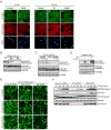
Fig. 1. Manipulating the expression of Bα and Bδ in Xenopus embryos produces distinct phenotypes
(A) Xenopus embryos were injected with either control GFP mRNA, Flag-tagged mouse Bδ mRNA (Bδ), a morpholino control (MoC) or a specific morpholino against Xenopus Bα (MoBα) or Bδ (MoBδ) at the one-cell stage, and fixed when control embryos had reached either early gastrula or tailbud (stage 25). Representative embryos are shown, arrows indicate anterior. The anterior regions of embryos are magnified below. Note the lack of head structures in Bδ-injected embryos and MoBα-injected embryos. (B) Embryos were injected as in A with the indicated mRNAs and morpholinos. The effect of MoBδ or MoBα could be rescued by coinjection with the cognate mRNA (mouse Bδ or Bα). Percentages of embryos showing wild type phenotype when control-injected embryos had reached stage 22 are given. Arrows indicate anterior.
Fig. 2
Fig. 2. Manipulating the expression of Bα and Bδ in Xenopus embryos has opposing effects on Activin/Nodal target gene expression
(A) In situ hybridisation of gastrula-stage embryos injected with either a morpholino control (MoC) or a specific morpholino against Xenopus Bα (MoBα) or Bδ (MoBδ) or with Flag-tagged mouse Bδ mRNA (Bδ) at the one-cell stage. The probes used were against gsc or Xbra. Staining was visualised with BM purple. Note the increased staining for both Xbra and Gsc in Bδ morphants, and decreased staining in Bα morphants and embryos overexpressing Bδ. (B) Analysis of gene expression by q-PCR. Total RNA was isolated from stage 10.5 embryos that had been injected at the one-cell stage with either morpholino control, or morpholinos against Bα or Bδ. Expression levels are normalised to ornithine decarboxylase (ODC).
Fig. 3
Fig. 3. Bα and Bδ act on the Activin/Nodal signalling pathway in Xenopus
(A-C) One-cell embryos were injected with the indicated mRNAs (Bα, Bδ or EGFP-Smad2) and morpholinos (MoC, MoBα or MoBδ). When control embryos had reached stage 8, the animal pole was excised and incubated with or without Activin for 16 hrs, and visualised for the degree of elongation.
Fig. 4
Fig. 4. Overexpression of Drosophila Smad2 (Smox) can rescue the effects of overexpression of Drosophila B subunit, Twins in the wing
(A) Phenotypically wild type wing from +/Y; UAS-tws23; UAS-Smox8D3 male. (B) Small, blistered wing from A9-GAL4; UAS-tws23; + male. All wings from these males are smaller than wild type; approximately 80% were cupped and blistered with little or no evidence of veins. (C) Wing from A9-GAL4; +; UAS-Smox8D3 male. In this genotype, wing veins formed a delta at the margin, and additional wing vein material was often observed (arrowheads). (D) Phenotypically normal wing from A9-GAL4; UAS-tws23; UAS-Smox8D3 male. All veins terminated normally at the margin (arrows).
Fig. 5
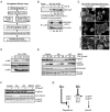
Fig. 5. Bα and Bδ exert differential effects on the level of phosphorylated Smad2
(A) Embryos were injected at the one-cell stage with morpholinos (Mo) against Bα or Bδ, or Bδ mRNA as indicated. Embryos were harvested at stage 10, fixed, dissected through the lip and analysed by immunofluorescence using anti-Smad2 and anti-β-Catenin antibodies. The nuclei were visualised with DAPI. Panels a and b show an area from the ventral vegetal region and panels c-e show an area from the dorsal vegetal region. (B) Embryos remained uninjected (ui) or were injected with two doses of mouse Bα mRNA or mouse Bδ mRNA, cultured until control embryos reached stage 9 and analysed by immunoblotting with anti-phospho-Smad2, Smad2/3, or phospho-ERK (pERK) antibodies. (C) Embryos were injected with distinct morpholinos (labelled 1 or 2) targeting Bδ, or Bα respectively, or with a control morpholino and analysed as in (B). (D) Animal caps from stage 8 embryos were incubated with or without okadaic acid (OA, 25 nM) for 1 hr, treated with or without Activin for 20 min and processed for immunoblotting. (E) HeLa EGFP-Smad2 cells were transfected with either an siRNA SMARTpool control or a human Bα- or Bδ-specific SMARTpool. Cells were incubated with TGF-β for the times indicated, fixed and visualised by confocal microscopy. (F) HeLa EGFP-Smad2 cells were transfected as in E and incubated with TGF-β for the times indicated. Samples were analysed by Western blotting with anti-phospho-Smad2, anti-Smad2/3 and anti-pan B subunit antibodies.
Fig. 6
Fig. 6. Bα and Bδ do not act directly on phosphorylated Smad2
(A) Outline of the experimental procedure to isolate Bα- and Bδ-containing active PP2A holocomplexes and to perform phosphatase assays. (B) Silver-stained gel showing the composition of complexes isolated by Flag pulldown from HeLa cells transfected with the indicated Flag-tagged B subunits. The components of the complex are indicated including the catalytic subunit (PP2AC) and the structural subunit (PP2AA). *indicates that the Flag-Bδ overlies PP2AA. (C) Western blot analysis of immunopurified complexes showing the presence of appropriate B or B′δ (PPP2R5D) subunit (Flag blot) and co-purified catalytic subunit (anti-PP2AC blot) for each complex. Phosphatase activity was assessed by a colorimetric assay using a phospho-peptide as substrate (bars). (D) PP2A complexes as in (C) were incubated with phospho-Smad2 immunopurified from TGF-β-induced HaCaT EGFP-Smad2 cells. The reactions were then analysed by immunoblotting with anti-pSmad2 and anti-Smad2/3 antibodies. All PP2A complexes tested failed to dephosphorylate phospho-Smad2. Bα- and Bδ-containing complexes dephosphorylated pS259 of immunoprecipitated HA-tagged Raf-1 (lower panels) (E) TGF-β treatment prior to immunopurification of the PP2A complexes does not affect the amount of co-purified catalytic subunit, nor the activity of the complexes in the colorimetric assay. (F) As in (D), but PP2A complexes were purified from untreated (-) or TGF-β-induced cells (+), as shown in (E). (G) Phosphorylated serines 245, 250 and 255 of Smad2 are not substrates for immunopurified Bα and Bδ complexes. Phosphatase complexes were immunopurified from either control cells (C) or cells expressing Flag-tagged Bα or Bδ as indicated, and incubated with either a Smad2/3 immunoprecipitate from TGF-β-induced HaCaT cells (upper panels) or, as a control, an immunopurified phosphorylated Raf substrate from HeLa cells expressing HA-Raf (lower panel). Samples were analysed by Western blotting using antibodies recognising Smad2 phosphorylated at residues S245, S250, S255, as well as anti-Smad2/3, anti-phospho Raf and anti-HA as indicated.
Fig. 7
Fig. 7. Bα regulates the basal level of the type I receptor and Bδ regulates its activity
(A) Outline of the experimental procedure to isolate Bα- and Bδ-containing active PP2A holocomplexes and to assay their ability to affect the kinase activity of ALK5. (B) The presence of neither PP2A complex affects the kinase activity of ALK5 in vitro. Endogenous ALK5 complexes immunopurified from untreated or TGF-β-treated HaCaT cells were incubated with recombinant Smad2 substrate in the absence or presence of B-subunit-specific PP2A complexes purified as in Figure 6. C-terminal Smad2 phosphorylation was detected by immunoblotting. The activity of the PP2A complexes was confirmed by their ability to dephosphorylate pS259 of Raf-1 (lower panel). (C) Knockdown of Bδ promotes ALK4 clustering. Animal caps from embryos expressing either HA-ALK4 mRNA alone (upper panels) or in combination with morpholino against Bδ (MoBδ, middle panels) were incubated for 1 hr in the presence or absence of Activin and stained with anti-HA antibody. HA-ALK4 clusters in response to Activin and in untreated embryos injected with MoBδ. Okadaic acid (OA) treatment (lower panels) also induces HA-ALK4 clustering and thus mimics Bδ knockdown. (D) Bα knockdown strongly decreases basal protein levels of ALK5. HaCaT cells were transfected with siRNAs and treated with TGF-β as indicated. Extracts were immunoblotted with antibodies against ALK5, phospho-Smad2, pan B-subunits and Smad2/3. (E) Bα knockdown has no effect on TβR-II levels. HaCaT cells were transfected with the indicated siRNAs. Extracts were immunoblotted with antibodies against TβR-II, pan B-subunits and Smad2/3. Prior to electrophoresis, extracts were treated ± PNGase F to remove N-linked sugars from TβR-II and visualise it more clearly. (F) Bα knockdown or Bδ overexpression decreases protein levels of HA-ALK4. Xenopus embryos were injected at the one-cell stage with HA-ALK4 and GFP mRNAs as well as with morpholinos or Bδ mRNA as indicated, cultured until uninjected embryos (Ui) had reached stage 9 and analysed by immunoblotting. (G) Model of the modulation of TGF-β/Activin/Nodal signalling by Bα and Bδ. Bα normally stabilises the type I receptors ALK4 and ALK5, and Bα knockdown promotes their basal degradation. Bδ normally restricts ligand-dependent activation of ALK4 and ALK5, and Bδ knockdown facilitates such activation. When overexpressed, Bδ additionally inihibits endogenous Bα by replacing it in the PP2A holoenzyme due to its higher affinity for the catalytic subunit.

References

    1. Adams DG, Coffee RL, Jr, Zhang H, Pelech S, Strack S, Wadzinski BE. Positive regulation of Raf1-MEK1/2-ERK1/2 signaling by protein serine/threonine phosphatase 2A holoenzymes. J Biol Chem. 2005;280:42644–54. - PubMed
    1. Bajpai R, Makhijani K, Rao PR, Shashidhara LS. Drosophila Twins regulates Armadillo levels in response to Wg/Wnt signal. Development. 2004;131:1007–16. - PubMed
    1. Bardwell VJ, Treisman R. The POZ domain: a conserved protein-protein interaction motif. Genes Dev. 1994;8:1664–77. - PubMed
    1. Batut J, Howell M, Hill CS. Kinesin-mediated transport of Smad2 is required for signaling in response to TGF-β ligands. Developmental Cell. 2007;12:261–274. - PubMed
    1. Bennett D, Alphey L. PP1 binds Sara and negatively regulates Dpp signaling in Drosophila melanogaster. Nat Genet. 2002;31:419–23. - PubMed

Publication types

MeSH terms

Substances