. 2008 Aug 19;105(33):11778-83.
doi: 10.1073/pnas.0711122105. Epub 2008 Aug 12.

Both cIAP1 and cIAP2 regulate TNFalpha-mediated NF-kappaB activation

Affiliations

Both cIAP1 and cIAP2 regulate TNFalpha-mediated NF-kappaB activation

D J Mahoney et al. Proc Natl Acad Sci U S A. .

Abstract

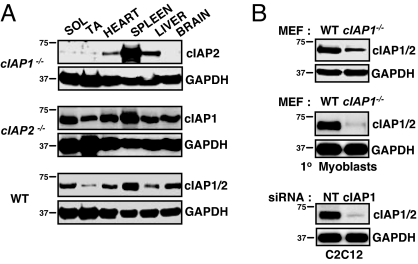
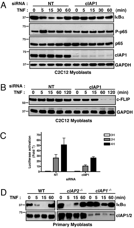
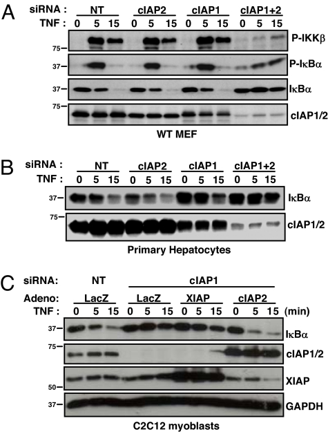
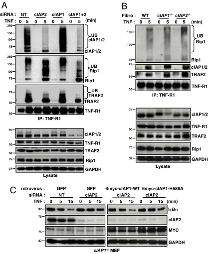
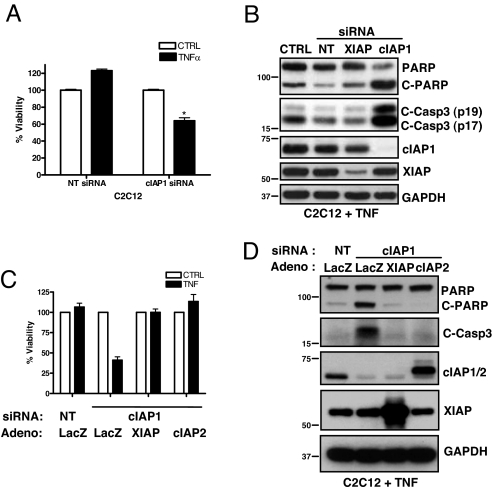
The cellular inhibitor of apoptosis 1 and 2 (cIAP1 and cIAP2) proteins have been implicated in the activation of NF-kappaB by TNFalpha; however, genetic deletion of either cIAP1 or 2 did not support a physiologically relevant role, perhaps because of functional redundancy. To address this, we used combined genetic and siRNA knockdown approaches and report that cIAP1 and 2 are indeed critical, yet redundant, regulators of NF-kappaB activation upon TNFalpha treatment. Whereas NF-kappaB was properly activated by TNFalpha in cultured and primary cells deficient in either cIAP1 or 2, removal of both cIAPs severely blunted its activation. After treatment with TNFalpha, cIAP1 and 2 were rapidly recruited to the TNF receptor 1, along with the adapter protein TNF receptor associated factor 2. Importantly, either cIAP1 or 2 was required for proper TNF receptor 1 signalosome function. In their combined absence, polyubiquitination of receptor interacting protein 1, an upstream event necessary for NF-kappaB signaling, was attenuated. As a result, phosphorylation of the inhibitor of kappaB kinase beta was diminished, and signal transduction was severely blunted. Consequently, cells missing both cIAP1 and 2 were sensitized to TNFalpha-mediated apoptosis. Collectively, these data demonstrate that either cIAP1 or 2 is required for proper Rip1 polyubiquitination and NF-kappaB activation upon TNFalpha treatment.


Conflict of interest statement

The authors declare no conflict of interest.

Figures

Fig. 1.
Fig. 1.
Tissue-specific elevation of cIAP2 in the cIAP1 null mouse. (A) Soleus (SOL), tibialis anterior (TA), heart, spleen, liver, and brain samples were taken from 3-month-old WT and cIAP1 and 2 null mice, homogenized, and protein lysates were immunoblotted for cIAP1 and 2 levels using our rabbit anti-rat IAP1 polyclonal antibody. (B) (Top) Primary mouse skeletal myoblasts were cultured, and cIAP1 and 2 levels were assessed by immunoblotting (MEFs were used as a control). (Bottom) Murine C2C12 myoblasts were treated with siRNA targeting cIAP1, and cIAP1 and 2 levels were assessed by immonoblotting.
Fig. 2.
Fig. 2.
TNFα-mediated NF-κB signaling in skeletal myoblasts depends on cIAP1. (A) C2C12 myoblasts were treated with siRNA for 24 h, treated with TNFα for the indicated times, and immunoblotted for members of the NF-κB signaling pathway. (B) C2C12 myoblasts were treated with siRNA for 24 h, treated with TNFα, and immunoblotted for c-FLIP. (C) C2C12 myoblasts stably expressing an NF-κB luciferase reporter construct were treated with siRNA for 24 h, followed by TNFα for the indicated amounts of time before luciferase activity was measured. Data are expressed mean fold change ± SD, n = 4. (D) Primary skeletal myoblasts were extracted, cultured for 24 h, and treated with TNFα. Protein lysates were collected and immunoblotted for IκBα.
Fig. 3.
Fig. 3.
cIAP1 and 2 redundantly regulate TNFα-mediated NF-κB activation in MEF cells and hepatocytes. (A) MEFs were treated with siRNA for 24 h, protein lysates extracted at the indicated times after TNFα treatment, and immunoblots were performed. (B) Primary hepatocytes were extracted from WT mice, treated with siRNA for 24 h, treated with TNFα for the indicated times, and immunoblotted. (C) C2C12 myoblasts were treated with siRNA and 4 h later infected with adenovirus (Adeno). Twenty hours later, cells were treated with TNFα, protein lysates collected, and immunoblots were performed.
Fig. 4.
Fig. 4.
Both cIAP1 and 2 are recruited to TNF-R1 upon TNFα treatment and are required for Rip1 polyubiquitination. (A) WT MEFs were treated with siRNA for 24 h, and endogenous TNF-R1 was immunoprecipitated before and 5 min after TNFα treatment and immunoblotted with the indicated antibodies. (B) Primary fibroblasts were cultured and treated with TNFα. TNF-R1 was immunoprecipitated, and immunoblots were performed as outlined. (C) cIAP1−/− MEFs were treated with siRNA and 8 h later infected with retrovirus. Forty-eight hours after siRNA treatment, cells were treated with TNFα and immunoblots were performed.
Fig. 5.
Fig. 5.
Either cIAP1 or 2 is required to protect against TNFα-induced apoptosis. (A) C2C12 myoblasts were treated with siRNA for 24 h, followed by TNFα treatment for 24 h, and cell viability was assessed. Data are expressed as % viability ± SD relative to no TNF treatment controls (set at 100%), n = 4 per condition. (B) C2C12 myoblasts treated with siRNA (24 h) followed by TNFα (24 h) were immunoblotted for markers of caspase activation. (C) C2C12 myoblasts were treated with siRNA and 4 h later infected with adenovirus (Adeno). After 24 h of knockdown, cells were treated with TNFα for 24 h, and cell viability was assessed (as above). (D) C2C12 cells were treated as in C, and protein lysates were immunoblotted for the indicated proteins.

Similar articles

Cited by

References

    1. Liston P, et al. Suppression of apoptosis in mammalian cells by NAIP and a related family of IAP genes. Nature. 1996;379:349–353. - PubMed
    1. Roy N, Deveraux QL, Takahashi R, Salvesen GS, Reed JC. The c-IAP-1 and c-IAP-2 proteins are direct inhibitors of specific caspases. EMBO J. 1997;16:6914–6925. - PMC - PubMed
    1. Samuel T, et al. Distinct BIR domains of cIAP1 mediate binding to and ubiquitination of tumor necrosis factor receptor-associated factor 2 and second mitochondrial activator of caspases. J Biol Chem. 2006;281:1080–1090. - PubMed
    1. Yang Y, Fang S, Jensen JP, Weissman AM, Ashwell JD. Ubiquitin protein ligase activity of IAPs and their degradation in proteasomes in response to apoptotic stimuli. Science. 2000;288:874–877. - PubMed
    1. Eckelman BP, Salvesen GS. The human anti-apoptotic proteins cIAP1 and cIAP2 bind but do not inhibit caspases. J Biol Chem. 2006;281:3254–3260. - PubMed

Publication types

MeSH terms

Substances