. 2008 Aug 19;105(33):11613-8.
doi: 10.1073/pnas.0801763105. Epub 2008 Aug 12.

The effect of particle design on cellular internalization pathways

Affiliations

The effect of particle design on cellular internalization pathways

Stephanie E A Gratton et al. Proc Natl Acad Sci U S A. .

Abstract

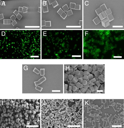
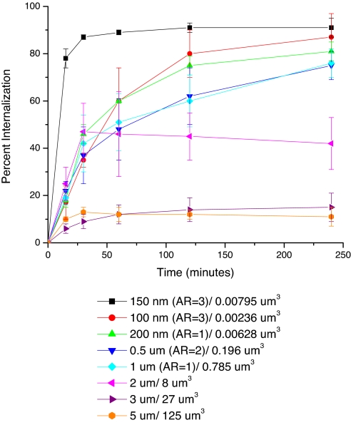
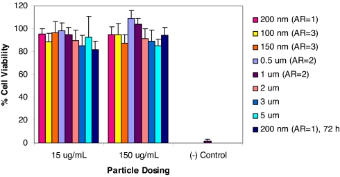
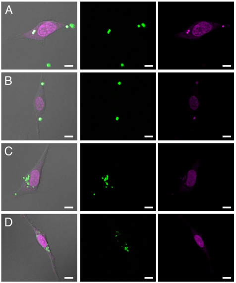
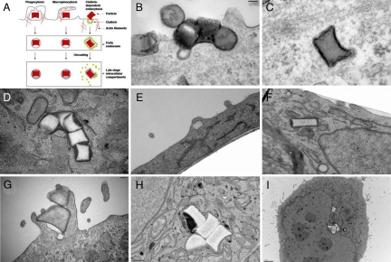
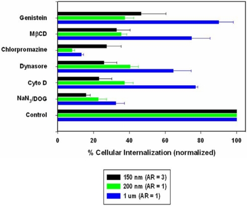
The interaction of particles with cells is known to be strongly influenced by particle size, but little is known about the interdependent role that size, shape, and surface chemistry have on cellular internalization and intracellular trafficking. We report on the internalization of specially designed, monodisperse hydrogel particles into HeLa cells as a function of size, shape, and surface charge. We employ a top-down particle fabrication technique called PRINT that is able to generate uniform populations of organic micro- and nanoparticles with complete control of size, shape, and surface chemistry. Evidence of particle internalization was obtained by using conventional biological techniques and transmission electron microscopy. These findings suggest that HeLa cells readily internalize nonspherical particles with dimensions as large as 3 mum by using several different mechanisms of endocytosis. Moreover, it was found that rod-like particles enjoy an appreciable advantage when it comes to internalization rates, reminiscent of the advantage that many rod-like bacteria have for internalization in nonphagocytic cells.


Conflict of interest statement

Conflict of interest statement: The research reported in this paper received partial financial support from a venture capital-backed company that Dr. Joseph M. DeSimone cofounded, Liquidia Technologies (www.liquidia.com). Currently he is on the board of directors and has personal financial interests in Liquidia Technologies.

Figures

Fig. 1.
Fig. 1.
Micrographs of PRINT particles varying in both size and shape. The particle composition for these experiments was 67 wt % trimethyloylpropane ethoxylate triacrylate (MW = 428 g/mol), 20 wt % poly(ethylene glycol) monomethylether monomethacrylate (MW = 1,000 g/mol), 10 wt % 2-aminoethylmethacrylate hydrochloride (AEM·HCl), 2 wt % fluorescein-O-acrylate, and 1 wt % 2,2-diethoxyacetophenone. (A–C) Scanning electron micrograph of the cubic series of particles [diameters equal to 2 μm (A), 3 μm (B), and 5 μm (C)]. (D–F) Fluorescence micrographs of the cubic series of particles [diameters equal to 2 μm (D), 3 μm (E), and 5 μm (F)]. (G and H) Scanning electron micrographs of the cylindrical series of microparticles having the same height (1 μm), but varying diameters [diameter = 0.5 μm (G) and 1 μm (H)]. (I–K) Scanning electron micrographs of the series of cylindrical nanoparticles [diameter = 200 nm, height = 200 nm (I); diameter = 100 nm, height = 300 nm (J); diameter = 150 nm, height = 450 nm (K)]. (Scale bars: A–F, 20 μm; G–K, 1 μm.)
Fig. 2.
Fig. 2.
Internalization profile of PRINT particles with HeLa cells over a 4-h incubation period at 37°C. Legend depicts the particle diameter per particle volume.
Fig. 3.
Fig. 3.
MTS assay showing the cytotoxicity of all particles under investigation. All experiments were carried out with a 4-h incubation with HeLa cells, except the final bar, where the 200-nm particles were tested for cytotoxicity to 72 h.
Fig. 4.
Fig. 4.
Confocal laser scanning microscopy images of HeLa cells after a 1-h incubation period at 37°C with 3-μm cubes (A), 2-μm cubes (B), 1-μm (AR = 1) cylinders (C), and 200-nm (AR = 1) cylindrical particles (D). (scale bar: 10 μm.)
Fig. 5.
Fig. 5.
Transmission electron microscopy images of HeLa cells at 37°C (incubation times in parentheses). (A) Illustration depicting the major pathways of cellular internalization used by PRINT particles. (B–D) With 200 nm (AR = 1) cylindrical particles (AR = 1) (B and C, 15 min; D, 4 h). (E and F) With 150 nm (AR = 3) cylindrical particles (E and F, 1 h). (G–I) With 1-μm (AR = 1) cylindrical particles (G, 1 h; H and I, 4 h).
Fig. 6.
Fig. 6.
Probing the mechanisms of cellular internalization by using inhibitors of endocytosis. HeLa cells were incubated with the indicated inhibitors in the graph as outlined in the experimental methods. Percent internalization was normalized to particle internalization in the absence of inhibitors.

References

    1. Cossart P. Subversion of the mammalian cell cytoskeleton by invasive bacteria. J Clin Invest. 1997;99:2307–2311. - PMC - PubMed
    1. Lukyanov AN, Elbayoumi TA, Chakilam AR, Torchilin VP. Tumor-targeted liposomes: doxorubicin-loaded long-circulating liposomes modified with anti-cancer antibody. J Controlled Release. 2004;100:135–144. - PubMed
    1. Torchilin VP. Targeted polymeric micelles for delivery of poorly soluble drugs. Cell Mol Life Sci. 2004;61:2549–2559. - PMC - PubMed
    1. Kabanov AV, Felgner PL, Seymour LW. Self-assembling Complexes for Gene Delivery. From Laboratory to Clinical Trial. Chichester: Wiley; 1998.
    1. Sarciaux JM, Acar L, Sado PA. Using microemulsion formulations for oral-drug delivery of therapeutic peptides. Int J Pharm. 1995;120:127–136.

Publication types