Prognostic significance of imaging contrast enhancement for WHO grade II gliomas
- PMID: 18697954
- PMCID: PMC2718989
- DOI: 10.1215/15228517-2008-066
Prognostic significance of imaging contrast enhancement for WHO grade II gliomas
Abstract
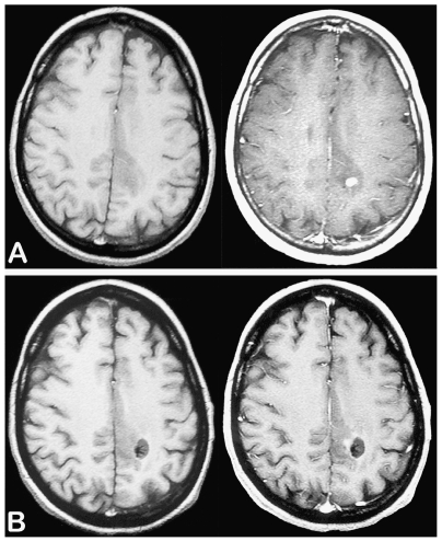
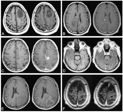
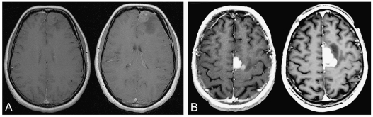
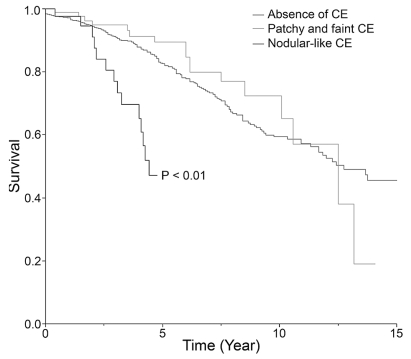
In this study, we investigated the prognostic value of MRI contrast enhancement (CE) at the time of histological diagnosis specifically in a selected population of WHO grade II gliomas. We reviewed 927 histologically proven WHO grade II gliomas for which contrast-enhanced MR images were available at the time of histological diagnosis. CE patterns were classified into three categories: "patchy and faint," "nodular-like," and "ring-like." CE progression over time was recorded before oncological treatment on successive MR images, when available. CE was present in 143 cases (15.9%), with 93 patchy and faint, 50 nodular-like, and no ring-like patterns. CE areas were time progressive before oncological treatment in 35 of the 56 available cases (62.5%). Regardless of its pattern, the presence of CE was not significantly associated with a worsened prognosis (p = 0.415) by univariate analysis. Only the nodular-like pattern of CE (p < 0.01) and the time-progressive CE (p < 0.001) in the available subgroup proved to be statistically associated with survival since first oncological treatment. The present results show the necessity, in cases of WHO grade II gliomas, to study CE at the time of histological diagnosis and, whenever possible, to follow its progression over time before oncological treatment. Nodular-like CE and time-progressive CE are associated with a worsened prognosis, both suggesting malignant transformation, even though histopathological examination cannot initially disclose signs of malignancy in those areas.
Figures


Similar articles
-
Clinical Significance of Discrepancy between Arterial Spin Labeling Images and Contrast-enhanced Images in the Diagnosis of Brain Tumors.Magn Reson Med Sci. 2015;14(4):313-9. doi: 10.2463/mrms.2014-0083. Epub 2015 Jun 23. Magn Reson Med Sci. 2015. PMID: 26104074
-
Identifying the association between contrast enhancement pattern, surgical resection, and prognosis in anaplastic glioma patients.Neuroradiology. 2016 Apr;58(4):367-74. doi: 10.1007/s00234-016-1640-y. Epub 2016 Jan 21. Neuroradiology. 2016. PMID: 26795126
-
Microvascular leakage and contrast enhancement as prognostic factors for recurrence in unfavorable low-grade gliomas.J Neurooncol. 2010 Mar;97(1):81-8. doi: 10.1007/s11060-009-9992-3. Epub 2009 Aug 29. J Neurooncol. 2010. PMID: 19727561
-
Comparison between contrast-enhanced magnetic resonance imaging and technetium 99m glucohepatonic acid single photon emission computed tomography with histopathologic correlation in gliomas.J Comput Assist Tomogr. 2006 Sep-Oct;30(5):723-33. doi: 10.1097/01.rct.0000228154.58281.88. J Comput Assist Tomogr. 2006. PMID: 16954918 Clinical Trial.
-
Diagnosis and treatment in neuro-oncology: an oncological perspective.Br J Radiol. 2011 Dec;84 Spec No 2(Spec Iss 2):S82-9. doi: 10.1259/bjr/18061999. Br J Radiol. 2011. PMID: 22433832 Free PMC article. Review.
Cited by
-
Imaging growth as a predictor of grade of malignancy and aggressiveness of IDH-mutant and 1p/19q-codeleted oligodendrogliomas in adults.Neuro Oncol. 2020 Jul 7;22(7):993-1005. doi: 10.1093/neuonc/noaa022. Neuro Oncol. 2020. PMID: 32025725 Free PMC article.
-
Prognostic and clinical significance of contrast enhancement in WHO grade 2 oligodendrogliomas.J Neurooncol. 2025 Apr;172(2):481-490. doi: 10.1007/s11060-024-04929-3. Epub 2025 Jan 19. J Neurooncol. 2025. PMID: 39827421
-
Imaging growth and isocitrate dehydrogenase 1 mutation are independent predictors for diffuse low-grade gliomas.Neuro Oncol. 2014 Aug;16(8):1100-9. doi: 10.1093/neuonc/nou085. Epub 2014 May 20. Neuro Oncol. 2014. PMID: 24847087 Free PMC article.
-
Vorasidenib, a Dual Inhibitor of Mutant IDH1/2, in Recurrent or Progressive Glioma; Results of a First-in-Human Phase I Trial.Clin Cancer Res. 2021 Aug 15;27(16):4491-4499. doi: 10.1158/1078-0432.CCR-21-0611. Epub 2021 Jun 2. Clin Cancer Res. 2021. PMID: 34078652 Free PMC article. Clinical Trial.
-
Lower-grade gliomas surgery guided by GRPR-targeting PET/NIR dual-modality image probe: a prospective and single-arm clinical trial.Theranostics. 2024 Jan 1;14(2):819-829. doi: 10.7150/thno.91554. eCollection 2024. Theranostics. 2024. PMID: 38169486 Free PMC article. Clinical Trial.
References
-
- Kleihues P, Cavenee W. Tumours of the Nervous System, Pathology and Genetics Classification of Tumours. Lyon, France: International Agency for Research on Cancer Press; 2000.
-
- Cavaliere R, Lopes MB, Schiff D. Low-grade gliomas: an update on pathology and therapy. Lancet Neurol. 2005;4:760–770. - PubMed
-
- Lang FF, Gilbert MR. Diffusely infiltrative low-grade gliomas in adults. J Clin Oncol. 2006;24:1236–1245. - PubMed
-
- Wessels PH, Weber WE, Raven G, et al. Supratentorial grade II astrocytoma: biological features and clinical course. Lancet Neurol. 2003;2:395–403. - PubMed
-
- Mandonnet E, Delattre JY, Tanguy ML, et al. Continuous growth of mean tumor diameter in a subset of grade II gliomas. Ann Neurol. 2003;53:524–528. - PubMed
MeSH terms
Substances
LinkOut - more resources
Full Text Sources
Medical