. 2008 Aug 13;3(8):e2924.
doi: 10.1371/journal.pone.0002924.

Microarray analysis of human monocytes infected with Francisella tularensis identifies new targets of host response subversion

Affiliations

Microarray analysis of human monocytes infected with Francisella tularensis identifies new targets of host response subversion

Jonathan P Butchar et al. PLoS One. .

Abstract

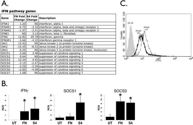
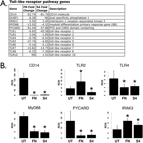
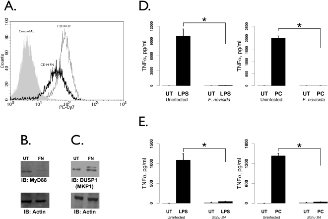
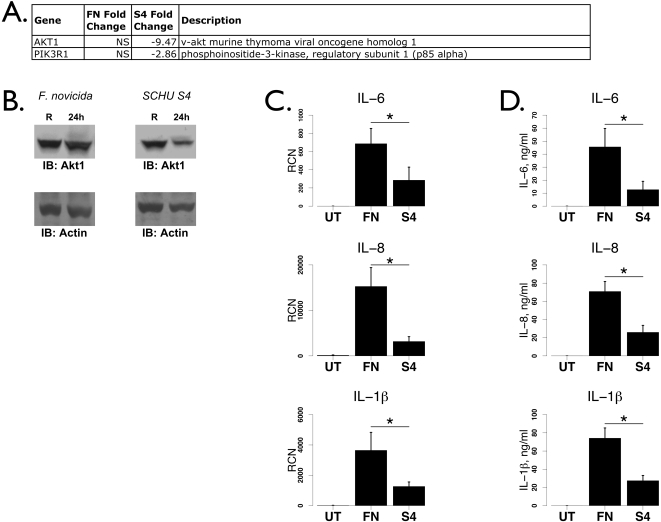
Francisella tularensis is a gram-negative facultative bacterium that causes the disease tularemia, even upon exposure to low numbers of bacteria. One critical characteristic of Francisella is its ability to dampen or subvert the host immune response. In order to help understand the mechanisms by which this occurs, we performed Affymetrix microarray analysis on transcripts from blood monocytes infected with the virulent Type A Schu S4 strain. Results showed that expression of several host response genes were reduced such as those associated with interferon signaling, Toll-like receptor signaling, autophagy and phagocytosis. When compared to microarrays from monocytes infected with the less virulent F. tularensis subsp. novicida, we found qualitative differences and also a general pattern of quantitatively reduced pro-inflammatory signaling pathway genes in the Schu S4 strain. Notably, the PI3K/Akt1 pathway appeared specifically down-regulated following Schu S4 infection and a concomitantly lower cytokine response was observed. This study identifies several new factors potentially important in host cell subversion by the virulent Type A F. tularensis that may serve as novel targets for drug discovery.


Conflict of interest statement

Competing Interests: The authors have declared that no competing interests exist.

Figures

Figure 1
Figure 1. Transcriptional responses to F. novicida and F. tularensis Schu S4.
A. Genes significantly different with an absolute fold change of 2 or greater after F. novicida or Schu S4 infection were pooled and used to create a heatmap. B. A Venn diagram of all significantly different genes that were up- or down-regulated twofold or more, with 6214 genes common to both strains, 3909 unique to FN, 3389 unique to S4 and 41163 counted as not different. FN: F. novicida (n = 4). S4: Schu S4 (n = 4). UT: untreated (n = 6).
Figure 2
Figure 2. Francisella-induced changes in IFNγ pathway-related genes.
A. List of IFN, Jak and SOCS genes regulated by Francisella infection (both F. novicida and Schu S4) from the microarray analysis. Fold changes of genes not statistically different are denoted “NS”. B. Real-time PCR of IFNγ, SOCS1 and SOCS3 from human PBM infected with either F. novicida (“FN”) or Schu S4 (“S4”) at 100 MOI for 24 hours (n = 8). “RCN” is Relative Copy Number for the Y axis. Asterisks denote statistical significance (p≤0.05) versus uninfected. C. Flow cytometry for the IFNγ receptor following F. novicida infection of human monocytes. The data are representative of three independent experiments.
Figure 3
Figure 3. Toll-like receptor pathway genes affected by Francisella.
A. List of TLR pathway genes from the microarray analysis. Fold changes of genes not statistically different are denoted “NS”. B. Real-time PCR analyses of CD14, TLR2, TLR4, MyD88, PYCARD and IRAK-M from human PBM infected with either F. novicida (“FN”) or Schu S4 (“S4”) at an MOI of 100 for 24 hours (n = 8). “RCN” is Relative Copy Number for the Y axis. Asterisks denote statistical significance (p≤0.05) versus uninfected.
Figure 4
Figure 4. Hyporesponsiveness of TLR signaling after Francisella infection.
A. Flow cytometry of CD14 in infected versus uninfected monocytes, gating on live cells by scatter. B and C. Western blot of MyD88 (B) and MKP1 / DUSP1 (C) after F. novicida infection of THP-1 cells. Actin reprobes (bottom panels) show equal loading. D and E. THP-1 cells were first infected with F. novicida (D) or Schu S4 (E) for 14 hours or left uninfected. Uninfected and infected cells were then treated with gentamicin at 50 µg/ml to kill extracellular bacteria and restimulated with LPS (500 ng/ml, D and E left panels) or Pam3CSK (100 ng/ml, D and E right panels) for 8 hours. Cell supernatants were analyzed for TNFα by ELISA. Values from three independent infections were analyzed by student's t-test. Asterisks denote statistical significance (p≤0.05).
Figure 5
Figure 5. Dampened cytokine response in Schu S4-infected monocytes.
A. Table of PI3K and Akt response genes from the microarray analysis. Genes not significantly different are denoted with “NS” in the Fold Change columns. B. Western blots for Akt1 after F. novicida (left) and Schu S4 (right) infection of human monocytes. Actin reprobes showed equivalent loading (bottom panels). The data are representative of three independent observations. C. Real-time PCR analysis of IL-8, IL-6 and IL-1β transcripts after infection of human peripheral blood moncytes by F. novicida (“FN”) and Schu S4 (“S4”) (n = 8). “RCN” is Relative Copy Number for the Y axis. D. ELISAs showing IL-8, IL-6 and IL-1β after infection by F. novicida and Schu S4 in human monocytes (n = 4). Data were analyzed by student's t-test. Asterisks denote statistical significance (p≤0.05).

References

    1. Proctor R, White J, Ayala E, Canonico P. Phagocytosis of Francisella tularensis by Rhesus monkey peripheral leukocytes. Infect Immun. 1975;11:146–151. - PMC - PubMed
    1. Lofgren S, Tarnvik A, Bloom G, Sjoberg W. Phagocytosis and killing of Francisella tularensis by human polymorphonuclear leukocytes. Infect Immun. 1983;39:715–720. - PMC - PubMed
    1. Hall J, Craven R, Fuller J, Pickles R, Kawula T. Francisella tularensis replicates within alveolar type II epithelial cells in vitro and in vivo following inhalation. Infect Immun. 2007;75:1034–1039. - PMC - PubMed
    1. Shaughnessy LM, Swanson JA. The role of the activated macrophage in clearing Listeria monocytogenes infection. Front Biosci. 2007;12:2683–2692. - PMC - PubMed
    1. Clemens D, Lee B, Horwitz M. Virulent and avirulent strains of Francisella tularensis prevent acidification and maturation of their phagosomes and escape into the cytoplasm in human macrophages. Infect Immun. 2004;72:3204–3217. - PMC - PubMed

Publication types