. 2008 Aug 13;3(8):e2952.
doi: 10.1371/journal.pone.0002952.

Suppression of HIV-specific and allogeneic T cell activation by human regulatory T cells is dependent on the strength of signals

Affiliations

Suppression of HIV-specific and allogeneic T cell activation by human regulatory T cells is dependent on the strength of signals

Amanda K Antons et al. PLoS One. .

Abstract

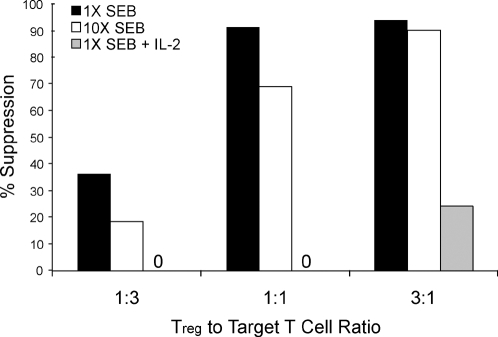
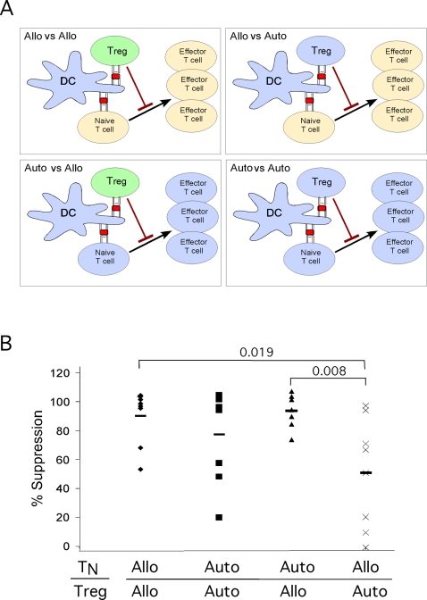
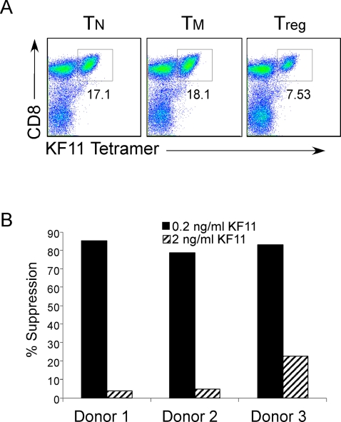
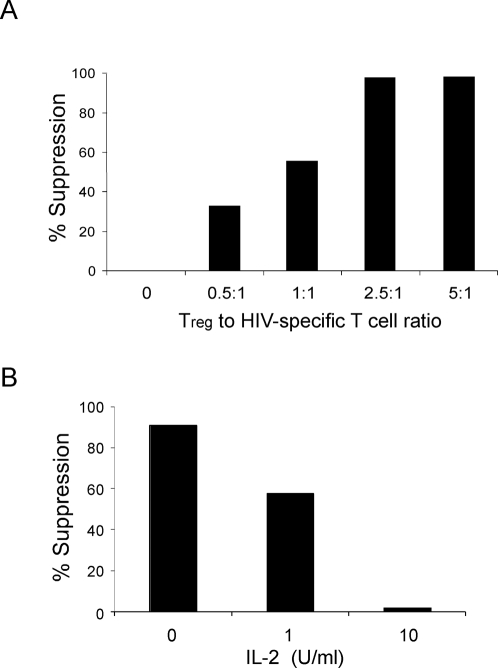
Regulatory T cells (Tregs) suppress immune responses against both self and non-self antigens. Tregs require activation through the T cell receptor (TCR) and IL-2 to exert their suppressive functions. However, how strength of TCR signals modulate the potency of Treg-mediated suppression of antigen-specific T cell activation remain unclear. We found that both strength of TCR signals and ratios of Tregs to target cells, either through superantigen, allogeneic antigens or HIV-specific peptides, modified the suppressive ability of Tregs. While human Tregs were able to mediate suppression in the presence of only autologous antigen-presenting cells, this was much less efficient as compared to when Tregs were activated by allogeneic dendritic cells. In another physiologically relevant system, we show that the strength of peptide stimulation, high frequency of responder CD8+ T cells or presence of high IL-2 can override the suppression of HIV-specific CD8+ T cells by Tregs. These findings suggest that ratios and TCR activation of human Tregs, are important parameters to overcome robust immune responses to pathogens or allogeneic antigens. Modulating the strength of T cell signals and selective enhancement or depletion of antigen-specific Tregs thus may have implications for designing potent vaccines and regulating immune responses during allogeneic transplantation and chronic infections.


Conflict of interest statement

Competing Interests: The authors have declared that no competing interests exist.

Figures

Figure 1
Figure 1. Strength of TCR activation and Treg numbers modulates level of suppression.
CD4+ naïve T cells were isolated and CFSE labeled. Unlabeled control TM or Tregs were added to the cultures at the target to Treg ratio as indicated. Cells were stimulated with 2 different concentrations of SEB, or in the presence of IL-2. Shown is percent suppression by the Tregs when normalized to TM or TN control. The % suppression on Y-axis is calculated as follows: We first determined the percent of CFSE labeled target cells dividing in response to the stimuli alone. We then determined the proliferative percentage of cells in the presence of Tregs or control effector T cells. The percent suppression was then calculated by percent reduction in proliferation of the target cells in the presence of effectors as compared to target cells alone. As an example, in one experiment 40% of the CFSE labeled T cells divided in response to SEB stimulation. In the presence of Tregs the same cells divided only 4%. Thus, we represent this as 90% suppression. Data shown are representative of three replicate experiments.
Figure 2
Figure 2. Treg mediated suppression of allogeneic T cell responses.
(A) Experimental setup allogeneic vs. autologous stimulation of CFSE labeled target cells (TN), and Tregs. (B) Conditions were designed as depicted in Figure 2A. Shown is percent suppression by the Tregs normalized to control T cells. Each point represents an individual experiment. in total 8 experiments separate experiments were performed. P values are shown above the brackets as calculated by Student's t Test.
Figure 3
Figure 3. Allogeneic Tregs are capable of suppressing antigen specific proliferation.
(A) To identify HIV-peptide specific CD8+ T cells PBMC cultures were stained with antibodies for CD3, CD4, CD8, and tetramer specific for HIV peptide, KF11. Plots shown are gated on CD3+ T cells, and tetramer positive cells were also CD8+. (B) Tregs or control T cells were isolated from HIV-negative individuals. PBMC from HIV+ individuals were CFSE labeled and mixed with either HIV-negative allogeneic T or Tregs, and stimulated with 0.2 or 2 ng/ml KF11 HIV peptide. Cells were stained with KF11 Tetramer, and level of proliferation was assessed d5 post activation. Shown is percent suppression by Tregs as described in Figure 1. Data is representative of 5 separate experiments.
Figure 4
Figure 4. Cell ratios and IL-2 effect Treg suppression of HIV specific proliferation.
(A) Tregs or control TN cells were isolated from HIV-negative individuals. PBMC from HIV+ were CFSE labeled and stimulated as indicated in Figure 3B. Tregs or control T cells were added at the ratio indicated. Shown is percent suppression by Tregs as described (Figure 1 legend). (B) HIV-positive PBMC was stimulated with KF11 peptide for 5 days in the presence of allogeneic Tregs and increasing concentrations of IL-2. Data is representative of three separate experiments.
Figure 5
Figure 5. Depletion of Tregs does not result in increased KF11-specific proliferation.
(A) PBMC from HIV+ individuals were stained with antibodies directed against CD4 and CD25. CD4+CD25hi cells were removed from the PBMC by sorting CD4+CD25hi T cells using FACS. Total or CD4+CD25hi depleted, or PBMC were then CFSE labeled and stimulated with KF11 peptide. Cells were stained as in Figure 3A, and percentage of KF11-specific CD8+ T cells that proliferated is shown for 3 representative donors. In conditions that CD4+CD25hi cells were added back to cultures, they were added at a ratio of at least 3∶1 CD4+CD25hi to labeled PBMC. (B) Treg, TN or TM cells were isolated from purified CD4+ T cells from HIV+ individual, based on expression of CD45RO and CD25. TN cells from the same donor were CFSE labeled, and unlabeled TN, TM or Tregs were added at a ratio of 1∶1 target to effector T cells. Cultures were stimulated with SEB pulsed DCs, and fixed on day 4–5 post activation. Histogram of CFSE labeled target cells are displayed, and the percentage of cells that have proliferated as shown in gates. Representative is one out of three experiments.

References

    1. Oswald-Richter K, Grill SM, Shariat N, Leelawong M, Sundrud MS, et al. HIV infection of naturally occurring and genetically reprogrammed human regulatory T-cells. PLoS Biol. 2004;2:E198. - PMC - PubMed
    1. Liu W, Putnam AL, Xu-Yu Z, Szot GL, Lee MR, et al. CD127 expression inversely correlates with FoxP3 and suppressive function of human CD4+ T reg cells. J Exp Med. 2006;203:1701–1711. - PMC - PubMed
    1. Banham AH. Cell-surface IL-7 receptor expression facilitates the purification of FOXP3(+) regulatory T cells. Trends Immunol. 2006;27:541–544. - PubMed
    1. Shevach EM. CD4+ CD25+ suppressor T cells: more questions than answers. Nat Rev Immunol. 2002;2:389–400. - PubMed
    1. Thornton AM, Shevach EM. Suppressor effector function of CD4+CD25+ immunoregulatory T cells is antigen nonspecific. J Immunol. 2000;164:183–190. - PubMed

Publication types

MeSH terms