

Analysis of the Prader-Willi syndrome chromosome region using quantitative microsphere hybridization (QMH) array
- PMID: 18698613
- PMCID: PMC5438262
- DOI: 10.1002/ajmg.a.32459
Analysis of the Prader-Willi syndrome chromosome region using quantitative microsphere hybridization (QMH) array
Abstract
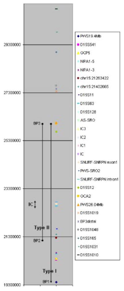
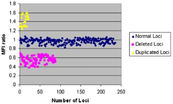
We previously developed a novel quantitative microsphere suspension hybridization (QMH) assay for high-throughput determination of genomic copy number by direct hybridization of unique sequence probes to genomic DNA followed by flow cytometric analysis. Herein, we describe the first clinical application of this assay examining the Prader-Willi syndrome (PWS) chromosome region at 15q11-13. We designed 30 unique sequence test probes (approximately 60 nucleotides each) spanning 11.37 Mb of chromosome 15q11.2-q13.3 and a disomic reference probe (Actin Beta, chromosome 7p22.1), conjugated to spectrally distinct polystyrene microsphere levels. All probes were hybridized to biotin-labeled genomic DNA in multiplex QMH reactions, and hybridization was detected using phycoerythrin-labeled streptavidin and analyzed by dual-laser flow cytometry. Copy number differences were distinguished by comparing mean fluorescence intensities (MFI) of the test probes to the reference probe in 20 individuals with PWS and six controls. The mean MFI ratio for deleted loci was 0.56 +/- 0.09 (n = 88) as compared to the MFI ratios for normal loci, 0.96 +/- 0.06 (n = 236), and duplicated loci, 1.44 +/- 0.10 (n = 22). A multiplex QMH assay could readily distinguish type I from type II deletions in PWS subjects, as well as small (approximately 4.3 kb) imprinting center (IC) deletions, with no overlap in MFI values compared with normal loci. Using this diagnostic QMH assay, the precise deleted genomic interval could be ascertained in all PWS subjects examined in the present study.
Copyright 2008 Wiley-Liss, Inc.
Figures
References
-
- Bejjani BA, Shaffer LG. A cytogeneticist’s perspective on genomic microarrays. Hum Reprod Update. 2004;10:221–226. - PubMed
-
- Bettio D, Rizzi N, Giardino D, Grugni G, Briscioli V, Selicorni A, Carnevale F, Larizza L. FISH analysis in Prader-Willi and Angelman syndrome patients. Am J Med Genet. 1995;56:224–228. - PubMed
MeSH terms
Substances
Grants and funding
LinkOut - more resources
Full Text Sources
Medical
Miscellaneous
