

Evidence of an inflammatory-like response in non-normally pigmented tissues of two scleractinian corals
- PMID: 18700208
- PMCID: PMC2605814
- DOI: 10.1098/rspb.2008.0335
Evidence of an inflammatory-like response in non-normally pigmented tissues of two scleractinian corals
Abstract
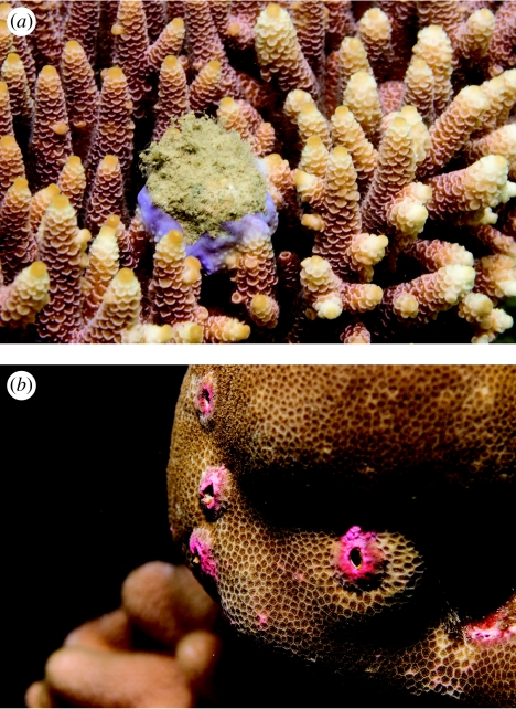
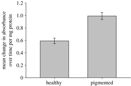
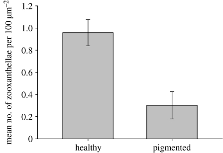
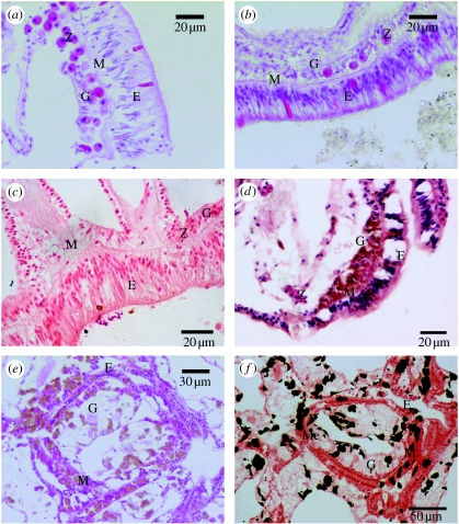
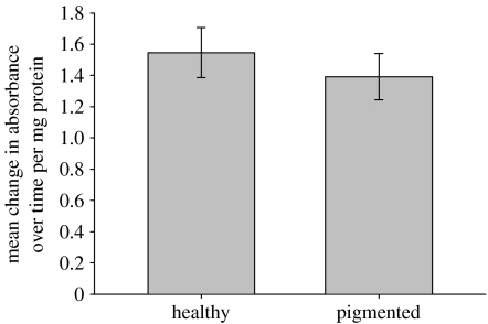
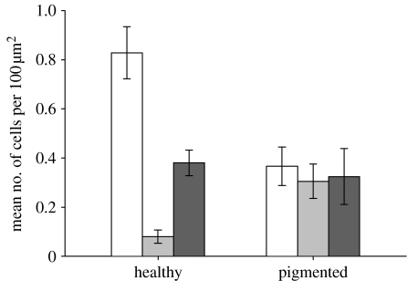
Increasing evidence of links between climate change, anthropogenic stress and coral disease underscores the importance of understanding the mechanisms by which reef-building corals resist infection and recover from injury. Cellular inflammation and melanin-producing signalling pathway are two mechanisms employed by invertebrates to remove foreign organisms such as pathogens, but they have not been recorded previously in scleractinian corals. This study demonstrates the presence of the phenoloxidase (PO) activating melanin pathway in two species of coral, Acropora millepora and a massive species of Porites, which both develop local pigmentation in response to interactions with a variety of organisms. L-DOPA (3-(3,4-dihydroxyphenyl)-L-alanine) substrate-based enzyme activation assays demonstrated PO activity in healthy tissues of both species and upregulation in pigmented tissues of A. millepora. Histological staining conclusively identified the presence of melanin in Porites tissues. These results demonstrate that the PO pathway is active in both coral species. Moreover, the upregulation of PO activity in areas of non-normal pigmentation in A. millepora and increased melanin production in pigmented Porites tissues suggest the presence of a generalized defence response to localized stress. Interspecific differences in the usage of pathways involved in innate immunity may underlie the comparative success of massive Porites sp. as long-lived stress tolerators.
Figures
Similar articles
-
A comparative study of phenoloxidase activity in diseased and bleached colonies of the coral Acropora millepora.Dev Comp Immunol. 2011 Oct;35(10):1098-101. doi: 10.1016/j.dci.2011.04.001. Epub 2011 Apr 19. Dev Comp Immunol. 2011. PMID: 21527282
-
The presence of multiple phenoloxidases in Caribbean reef-building corals.Comp Biochem Physiol A Mol Integr Physiol. 2011 Aug;159(4):372-8. doi: 10.1016/j.cbpa.2011.03.029. Epub 2011 Apr 8. Comp Biochem Physiol A Mol Integr Physiol. 2011. PMID: 21497202
-
Levels of immunity parameters underpin bleaching and disease susceptibility of reef corals.FASEB J. 2010 Jun;24(6):1935-46. doi: 10.1096/fj.09-152447. Epub 2010 Feb 2. FASEB J. 2010. PMID: 20124432
-
Biological and remote sensing perspectives of pigmentation in coral reef organisms.Adv Mar Biol. 2002;43:277-317. doi: 10.1016/s0065-2881(02)43006-4. Adv Mar Biol. 2002. PMID: 12154614 Review.
-
The proPO-system: pros and cons for its role in invertebrate immunity.Trends Immunol. 2008 Jun;29(6):263-71. doi: 10.1016/j.it.2008.02.009. Epub 2008 May 3. Trends Immunol. 2008. PMID: 18457993 Review.
Cited by
-
Metabolomics of reef benthic interactions reveals a bioactive lipid involved in coral defence.Proc Biol Sci. 2016 Apr 27;283(1829):20160469. doi: 10.1098/rspb.2016.0469. Proc Biol Sci. 2016. PMID: 27122568 Free PMC article.
-
Hyperspectral sensing of disease stress in the Caribbean reef-building coral, Orbicella faveolata - perspectives for the field of coral disease monitoring.PLoS One. 2013 Dec 4;8(12):e81478. doi: 10.1371/journal.pone.0081478. eCollection 2013. PLoS One. 2013. PMID: 24324697 Free PMC article.
-
Thermal stress triggers broad Pocillopora damicornis transcriptomic remodeling, while Vibrio coralliilyticus infection induces a more targeted immuno-suppression response.PLoS One. 2014 Sep 26;9(9):e107672. doi: 10.1371/journal.pone.0107672. eCollection 2014. PLoS One. 2014. PMID: 25259845 Free PMC article.
-
Expression and Characterization of a Bright Far-red Fluorescent Protein from the Pink-Pigmented Tissues of Porites lobata.Mar Biotechnol (NY). 2020 Feb;22(1):67-80. doi: 10.1007/s10126-019-09931-9. Epub 2019 Dec 18. Mar Biotechnol (NY). 2020. PMID: 31853751
-
Before platelets: the production of platelet-activating factor during growth and stress in a basal marine organism.Proc Biol Sci. 2018 Aug 15;285(1884):20181307. doi: 10.1098/rspb.2018.1307. Proc Biol Sci. 2018. PMID: 30111600 Free PMC article.
References
-
- Aeby G.S. Corals in the genus Porites are susceptible to infection by a larval trematode. Coral Reefs. 2003;22:216. doi:10.1007/s00338-003-0310-9 - DOI
-
- Adema C.M, Van der Knaap W.P.W, Sminia T. Molluscan hemocytemediated cytotoxicity: the role of reactive oxygen intermediates. Rev. Aquat. Sci. 1991;4:201–223.
-
- Allam B, Ashton-Alcox K.A, Ford S.E. Haemocyte parameters associated with resistance to brown ring disease in Ruditapes spp. clams. Dev. Comp. Immunol. 2001;25:365–375. doi:10.1016/S0145-305X(00)00072-0 - DOI - PubMed
-
- Asokan R, Arumugam M, Mullainadhan P. Activation of prophenoloxidase in the plasma and haemocytes of the marine mussel, Perna viridis Linnaeus. Dev. Comp. Immunol. 1997;21:1–12. doi:10.1016/S0145-305X(97)00004-9 - DOI - PubMed
-
- Bayne C.J. Phagocytosis and non-self recognition in invertebrates. BioScience. 1990;40:723–731. doi:10.2307/1311504 - DOI
Publication types
MeSH terms
Substances
LinkOut - more resources
Full Text Sources
